 Movie
Movie Controller


+ Open data

- Basic information
| Entry | Database: PDB / ID: 7ptt | ||||||||||||
|---|---|---|---|---|---|---|---|---|---|---|---|---|---|
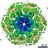
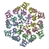
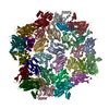
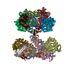
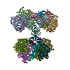
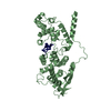
| Title | In-situ structure of hexameric S-layer protein | ||||||||||||
 Components Components | Cell surface glycoprotein | ||||||||||||
 Keywords Keywords | STRUCTURAL PROTEIN / S-layer csg | ||||||||||||
| Function / homology | Surface glycoprotein signal peptide / Major cell surface glycoprotein / PGF-CTERM archaeal protein-sorting signal / PGF-CTERM motif / S-layer / cell wall organization / plasma membrane / Cell surface glycoprotein Function and homology information Function and homology information | ||||||||||||
| Biological species |  Haloferax volcanii DS2 (archaea) Haloferax volcanii DS2 (archaea) | ||||||||||||
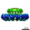
| Method | ELECTRON MICROSCOPY / subtomogram averaging / cryo EM / Resolution: 7.968 Å | ||||||||||||
 Authors Authors | von Kuegelgen, A. / Bharat, T.A.M. | ||||||||||||
| Funding support |  United Kingdom, 3items United Kingdom, 3items
| ||||||||||||
 Citation Citation | Journal: Cell Rep / Year: 2021 Title: Complete atomic structure of a native archaeal cell surface. Authors: Andriko von Kügelgen / Vikram Alva / Tanmay A M Bharat /  Abstract: Many prokaryotic cells are covered by an ordered, proteinaceous, sheet-like structure called a surface layer (S-layer). S-layer proteins (SLPs) are usually the highest copy number macromolecules in ...Many prokaryotic cells are covered by an ordered, proteinaceous, sheet-like structure called a surface layer (S-layer). S-layer proteins (SLPs) are usually the highest copy number macromolecules in prokaryotes, playing critical roles in cellular physiology such as blocking predators, scaffolding membranes, and facilitating environmental interactions. Using electron cryomicroscopy of two-dimensional sheets, we report the atomic structure of the S-layer from the archaeal model organism Haloferax volcanii. This S-layer consists of a hexagonal array of tightly interacting immunoglobulin-like domains, which are also found in SLPs across several classes of archaea. Cellular tomography reveal that the S-layer is nearly continuous on the cell surface, completed by pentameric defects in the hexagonal lattice. We further report the atomic structure of the SLP pentamer, which shows markedly different relative arrangements of SLP domains needed to complete the S-layer. Our structural data provide a framework for understanding cell surfaces of archaea at the atomic level. | ||||||||||||
| History |
|
- Structure visualization
| Movie |
Movie viewer |
|---|---|
| Structure viewer | Molecule: MolmilJmol/JSmol |
- Downloads & links
-Download
| PDBx/mmCIF format | 7ptt.cif.gz | 681.5 KB | Display | PDBx/mmCIF format |
|---|---|---|---|---|
| PDB format | pdb7ptt.ent.gz | 571.2 KB | Display | PDB format |
| PDBx/mmJSON format | 7ptt.json.gz | Tree view | PDBx/mmJSON format | |
| Others |  Other downloads Other downloads |
-Validation report
| Arichive directory | https://data.pdbj.org/pub/pdb/validation_reports/pt/7pttftp://data.pdbj.org/pub/pdb/validation_reports/pt/7ptt | HTTPS FTP |
|---|
-Related structure data
| Related structure data |  13637MC  7ptpC  7ptrC 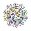 7ptuC M: map data used to model this data C: citing same article ( |
|---|---|
| Similar structure data |


-Links
 PDBj
PDBj- Assembly
| Deposited unit | 
|
|---|---|
| 1 |
|
-Components
| #1: Protein | Mass: 81755.602 Da / Num. of mol.: 6 / Source method: isolated from a natural source / Source: (natural) Haloferax volcanii DS2 (archaea) / Plasmid details: Allers et al 2004 / References: UniProt: P25062 |
|---|
-Experimental details
-Experiment
| Experiment | Method: ELECTRON MICROSCOPY |
|---|---|
| EM experiment | Aggregation state: CELL / 3D reconstruction method: subtomogram averaging |
- Sample preparation
| Component | Name: In-situ structure of hexameric S-layer of Haloferax volcanii Type: ORGANELLE OR CELLULAR COMPONENT Details: In-situ structure of hexameric S-layer of Haloferax volcanii Entity ID: all / Source: NATURAL | |||||||||||||||||||||||||||||||||||
|---|---|---|---|---|---|---|---|---|---|---|---|---|---|---|---|---|---|---|---|---|---|---|---|---|---|---|---|---|---|---|---|---|---|---|---|---|
| Molecular weight | Experimental value: NO | |||||||||||||||||||||||||||||||||||
| Source (natural) | Organism: Haloferax volcanii DS2 (archaea) / Cellular location: Cell surface | |||||||||||||||||||||||||||||||||||
| Buffer solution | pH: 7.5 / Details: 18 % (w/v) artificial sea water | |||||||||||||||||||||||||||||||||||
| Buffer component |
| |||||||||||||||||||||||||||||||||||
| Specimen | Embedding applied: NO / Shadowing applied: NO / Staining applied: NO / Vitrification applied: YES / Details: Haloferax volcanii vesicles | |||||||||||||||||||||||||||||||||||
| Specimen support | Details: 20 seconds, 15 mA / Grid material: COPPER/RHODIUM / Grid mesh size: 200 divisions/in. / Grid type: Quantifoil R2/2 | |||||||||||||||||||||||||||||||||||
| Vitrification | Instrument: FEI VITROBOT MARK IV / Cryogen name: ETHANE / Humidity: 100 % / Chamber temperature: 283.15 K Details: Vitrobot options: Blot time 2.0 seconds, Blot force -15,1, Wait time 0 seconds, Drain time 0.5 seconds |
- Electron microscopy imaging
| Experimental equipment |  Model: Titan Krios / Image courtesy: FEI Company |
|---|---|
| Microscopy | Model: FEI TITAN KRIOS / Details: SerialEM Hagen Scheme |
| Electron gun | Electron source:  FIELD EMISSION GUN / Accelerating voltage: 300 kV / Illumination mode: FLOOD BEAM FIELD EMISSION GUN / Accelerating voltage: 300 kV / Illumination mode: FLOOD BEAM |
| Electron lens | Mode: BRIGHT FIELD / Nominal magnification: 105000 X / Calibrated magnification: 105000 X / Nominal defocus max: 4000 nm / Nominal defocus min: 1000 nm / Calibrated defocus min: 1000 nm / Calibrated defocus max: 4000 nm / Cs: 2.7 mm / C2 aperture diameter: 50 µm / Alignment procedure: ZEMLIN TABLEAU |
| Specimen holder | Cryogen: NITROGEN / Specimen holder model: FEI TITAN KRIOS AUTOGRID HOLDER / Temperature (max): 70 K / Temperature (min): 70 K |
| Image recording | Average exposure time: 1 sec. / Electron dose: 2.9 e/Å2 / Detector mode: COUNTING / Film or detector model: GATAN K2 SUMMIT (4k x 4k) / Num. of grids imaged: 1 / Details: Dose symmetric tilt scheme (Hagen et al, JSB) |
| EM imaging optics | Energyfilter name: GIF Quantum LS / Chromatic aberration corrector: not used / Energyfilter slit width: 20 eV / Spherical aberration corrector: not used |
| Image scans | Width: 3838 / Height: 3710 |
- Processing
| Software | Name: PHENIX / Version: 1.19_4092: / Classification: refinement | ||||||||||||||||||||||||||||||||||||||||||||||||||
|---|---|---|---|---|---|---|---|---|---|---|---|---|---|---|---|---|---|---|---|---|---|---|---|---|---|---|---|---|---|---|---|---|---|---|---|---|---|---|---|---|---|---|---|---|---|---|---|---|---|---|---|
| EM software |
| ||||||||||||||||||||||||||||||||||||||||||||||||||
| CTF correction | Details: RELION subtomogram averaging (Bharat & Scheres 2016) Type: PHASE FLIPPING AND AMPLITUDE CORRECTION | ||||||||||||||||||||||||||||||||||||||||||||||||||
| Particle selection | Num. of particles selected: 1773652 Details: Particles were initially picking using the Laplacian-of gaussian algorithm implemented in RELION3.0 (Zivanov et al., 2018). Particles were extracted in 8x down-sampled in 50x50 pixel boxes ...Details: Particles were initially picking using the Laplacian-of gaussian algorithm implemented in RELION3.0 (Zivanov et al., 2018). Particles were extracted in 8x down-sampled in 50x50 pixel boxes and classified using reference-free 2D classification inside RELION3.0. | ||||||||||||||||||||||||||||||||||||||||||||||||||
| Symmetry | Point symmetry: C6 (6 fold cyclic) | ||||||||||||||||||||||||||||||||||||||||||||||||||
| 3D reconstruction | Resolution: 7.968 Å / Resolution method: FSC 0.143 CUT-OFF / Num. of particles: 53063 / Algorithm: FOURIER SPACE / Num. of class averages: 5 / Symmetry type: POINT | ||||||||||||||||||||||||||||||||||||||||||||||||||
| EM volume selection | Method: RELION / Details: RELION subtomogram averaging / Num. of tomograms: 127 / Num. of volumes extracted: 83713 / Reference model: Ab initio | ||||||||||||||||||||||||||||||||||||||||||||||||||
| Atomic model building | Protocol: RIGID BODY FIT / Space: REAL / Target criteria: Best Fit Details: Rigid body fit inside coot of D1-D6 domains and real space refinement with restraints of the original model obtained by single particle analysis in PHENIX. |
