 Movie
Movie Controller


+ Open data

- Basic information
| Entry | Database: PDB / ID: 7ori | ||||||
|---|---|---|---|---|---|---|---|
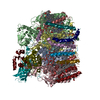
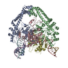
| Title | La Crosse virus polymerase at replication late-elongation stage | ||||||
 Components Components |
| ||||||
 Keywords Keywords | VIRAL PROTEIN / RNA-dependent RNA polymerase | ||||||
| Function / homology |  Function and homology information Function and homology informationhost cell endoplasmic reticulum / virion component / host cell endoplasmic reticulum-Golgi intermediate compartment / Hydrolases; Acting on ester bonds / host cell Golgi apparatus / RNA-directed RNA polymerase / viral RNA genome replication / nucleotide binding / hydrolase activity / RNA-directed RNA polymerase activity ...host cell endoplasmic reticulum / virion component / host cell endoplasmic reticulum-Golgi intermediate compartment / Hydrolases; Acting on ester bonds / host cell Golgi apparatus / RNA-directed RNA polymerase / viral RNA genome replication / nucleotide binding / hydrolase activity / RNA-directed RNA polymerase activity / DNA-templated transcription / RNA binding / metal ion binding Similarity search - Function | ||||||
| Biological species |  La Crosse orthobunyavirus La Crosse orthobunyavirus | ||||||
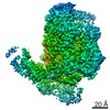
| Method | ELECTRON MICROSCOPY / single particle reconstruction / cryo EM / Resolution: 3.9 Å | ||||||
 Authors Authors | Arragain, B. / Durieux Trouilleton, Q. / Baudin, F. / Cusack, S. / Schoehn, G. / Malet, H. | ||||||
| Funding support |  France, 1items France, 1items
| ||||||
 Citation Citation | Journal: Nat Commun / Year: 2022 Title: Structural snapshots of La Crosse virus polymerase reveal the mechanisms underlying Peribunyaviridae replication and transcription. Authors: Benoît Arragain / Quentin Durieux Trouilleton / Florence Baudin / Jan Provaznik / Nayara Azevedo / Stephen Cusack / Guy Schoehn / Hélène Malet /  Abstract: Segmented negative-strand RNA bunyaviruses encode a multi-functional polymerase that performs genome replication and transcription. Here, we establish conditions for in vitro activity of La Crosse ...Segmented negative-strand RNA bunyaviruses encode a multi-functional polymerase that performs genome replication and transcription. Here, we establish conditions for in vitro activity of La Crosse virus polymerase and visualize its conformational dynamics by cryo-electron microscopy, unveiling the precise molecular mechanics underlying its essential activities. We find that replication initiation is coupled to distal duplex promoter formation, endonuclease movement, prime-and-realign loop extension and closure of the polymerase core that direct the template towards the active site. Transcription initiation depends on C-terminal region closure and endonuclease movements that prompt primer cleavage prior to primer entry in the active site. Product realignment after priming, observed in replication and transcription, is triggered by the prime-and-realign loop. Switch to elongation results in polymerase reorganization and core region opening to facilitate template-product duplex formation in the active site cavity. The uncovered detailed mechanics should be helpful for the future design of antivirals counteracting bunyaviral life threatening pathogens. | ||||||
| History |
|
- Structure visualization
| Movie |
Movie viewer |
|---|---|
| Structure viewer | Molecule: MolmilJmol/JSmol |
- Downloads & links
-Download
| PDBx/mmCIF format | 7ori.cif.gz | 402 KB | Display | PDBx/mmCIF format |
|---|---|---|---|---|
| PDB format | pdb7ori.ent.gz | 312.4 KB | Display | PDB format |
| PDBx/mmJSON format | 7ori.json.gz | Tree view | PDBx/mmJSON format | |
| Others |  Other downloads Other downloads |
-Validation report
| Arichive directory | https://data.pdbj.org/pub/pdb/validation_reports/or/7oriftp://data.pdbj.org/pub/pdb/validation_reports/or/7ori | HTTPS FTP |
|---|
-Related structure data
| Related structure data |  13038MC  7orjC  7orkC  7orlC  7ormC  7ornC  7oroC M: map data used to model this data C: citing same article ( |
|---|---|
| Similar structure data |


-Links
 PDBj
PDBj


- Assembly
| Deposited unit | 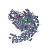
|
|---|---|
| 1 |
|
-Components
-RNA chain , 3 types, 3 molecules HTP
| #1: RNA chain | Mass: 5466.325 Da / Num. of mol.: 1 / Source method: obtained synthetically Details: mutated 5vRNA of La Crosse virus segment M Mutation of the nucleotides G2, U3, A9 and C10 of the 5 end into C2, G3, C9 and G10 Source: (synth.) La Crosse orthobunyavirus |
|---|---|
| #2: RNA chain | Mass: 9497.634 Da / Num. of mol.: 1 / Source method: obtained synthetically / Source: (synth.) La Crosse orthobunyavirus |
| #3: RNA chain | Mass: 9623.761 Da / Num. of mol.: 1 / Source method: isolated from a natural source Details: Product synthetized by La Crosse virus polymerase when incubated with its 5 RNA promoter, RNA template and nucleotides. Source: (natural) La Crosse orthobunyavirus |
-Protein , 1 types, 1 molecules A
| #4: Protein | Mass: 264751.062 Da / Num. of mol.: 1 / Mutation: H34K Source method: isolated from a genetically manipulated source Source: (gene. exp.) La Crosse orthobunyavirus / Gene: L segment / Cell line (production host): Hi 5 / Production host:  Trichoplusia ni (cabbage looper) / References: UniProt: A5HC98, RNA-directed RNA polymerase Trichoplusia ni (cabbage looper) / References: UniProt: A5HC98, RNA-directed RNA polymerase |
|---|
-Non-polymers , 3 types, 3 molecules 


| #5: Chemical | ChemComp-ZN /  Mass: 65.409 Da / Num. of mol.: 1 / Source method: obtained synthetically / Formula: Zn Mass: 65.409 Da / Num. of mol.: 1 / Source method: obtained synthetically / Formula: Zn |
|---|---|
| #6: Chemical | ChemComp-MG /  Mass: 24.305 Da / Num. of mol.: 1 / Source method: obtained synthetically / Formula: Mg Mass: 24.305 Da / Num. of mol.: 1 / Source method: obtained synthetically / Formula: Mg |
| #7: Chemical | ChemComp-POP /  Mass: 175.959 Da / Num. of mol.: 1 / Source method: obtained synthetically / Formula: H2O7P2 Mass: 175.959 Da / Num. of mol.: 1 / Source method: obtained synthetically / Formula: H2O7P2 |
-Details
| Has ligand of interest | N |
|---|
-Experimental details
-Experiment
| Experiment | Method: ELECTRON MICROSCOPY |
|---|---|
| EM experiment | Aggregation state: PARTICLE / 3D reconstruction method: single particle reconstruction |
- Sample preparation
| Component |
| ||||||||||||||||||||||||||||||||||||
|---|---|---|---|---|---|---|---|---|---|---|---|---|---|---|---|---|---|---|---|---|---|---|---|---|---|---|---|---|---|---|---|---|---|---|---|---|---|
| Molecular weight | Value: 0.291 MDa / Experimental value: NO | ||||||||||||||||||||||||||||||||||||
| Source (natural) |
| ||||||||||||||||||||||||||||||||||||
| Source (recombinant) |
| ||||||||||||||||||||||||||||||||||||
| Buffer solution | pH: 8 | ||||||||||||||||||||||||||||||||||||
| Buffer component |
| ||||||||||||||||||||||||||||||||||||
| Specimen | Conc.: 0.45 mg/ml / Embedding applied: NO / Shadowing applied: NO / Staining applied: NO / Vitrification applied: YES Details: 1.7 uM LACV-LCItag_H34K were sequentially incubated for 1h at 4degree with (i) 1.9 uM 5prime 1-17 BPm, (ii) 1.9 uM 3prime vRNA 1-30. LACV-LCItag_H34K bound to vRNAs was incubated with 100 uM ...Details: 1.7 uM LACV-LCItag_H34K were sequentially incubated for 1h at 4degree with (i) 1.9 uM 5prime 1-17 BPm, (ii) 1.9 uM 3prime vRNA 1-30. LACV-LCItag_H34K bound to vRNAs was incubated with 100 uM ATP/GTP/UTP/CTP and 5mM MgCl2 for 4h at 30degree | ||||||||||||||||||||||||||||||||||||
| Specimen support | Details: 25 mA / Grid material: GOLD / Grid mesh size: 300 divisions/in. / Grid type: UltrAuFoil R1.2/1.3 | ||||||||||||||||||||||||||||||||||||
| Vitrification | Instrument: FEI VITROBOT MARK IV / Cryogen name: ETHANE / Humidity: 100 % / Chamber temperature: 293 K |
- Electron microscopy imaging
| Microscopy | Model: TFS GLACIOS |
|---|---|
| Electron gun | Electron source:  FIELD EMISSION GUN / Accelerating voltage: 200 kV / Illumination mode: FLOOD BEAM FIELD EMISSION GUN / Accelerating voltage: 200 kV / Illumination mode: FLOOD BEAM |
| Electron lens | Mode: BRIGHT FIELD / Nominal magnification: 36000 X / Calibrated magnification: 36000 X / Nominal defocus max: 2000 nm / Nominal defocus min: 800 nm / Calibrated defocus min: 800 nm / Calibrated defocus max: 2000 nm / Cs: 2.7 mm / C2 aperture diameter: 100 µm / Alignment procedure: COMA FREE |
| Specimen holder | Cryogen: NITROGEN / Specimen holder model: FEI TITAN KRIOS AUTOGRID HOLDER / Temperature (max): 70 K / Temperature (min): 70 K |
| Image recording | Average exposure time: 6.6 sec. / Electron dose: 60 e/Å2 / Detector mode: COUNTING / Film or detector model: GATAN K2 SUMMIT (4k x 4k) / Num. of grids imaged: 1 / Num. of real images: 1848 |
| Image scans | Movie frames/image: 60 / Used frames/image: 3-50 |
- Processing
| EM software |
| |||||||||||||||||||||||||||||||||||||||||||||
|---|---|---|---|---|---|---|---|---|---|---|---|---|---|---|---|---|---|---|---|---|---|---|---|---|---|---|---|---|---|---|---|---|---|---|---|---|---|---|---|---|---|---|---|---|---|---|
| CTF correction | Type: PHASE FLIPPING AND AMPLITUDE CORRECTION | |||||||||||||||||||||||||||||||||||||||||||||
| Particle selection | Num. of particles selected: 1200554 | |||||||||||||||||||||||||||||||||||||||||||||
| Symmetry | Point symmetry: C1 (asymmetric) | |||||||||||||||||||||||||||||||||||||||||||||
| 3D reconstruction | Resolution: 3.9 Å / Resolution method: FSC 0.143 CUT-OFF / Num. of particles: 24151 / Algorithm: FOURIER SPACE / Symmetry type: POINT | |||||||||||||||||||||||||||||||||||||||||||||
| Atomic model building | B value: 109.75 / Protocol: AB INITIO MODEL | |||||||||||||||||||||||||||||||||||||||||||||
| Atomic model building | PDB-ID: 6Z8K Pdb chain-ID: A / Accession code: 6Z8K / Pdb chain residue range: 1-2263 / Source name: PDB / Type: experimental model |

