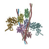
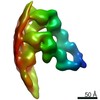
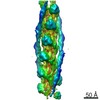
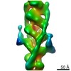
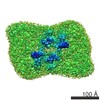
登録情報 データベース : PDB / ID : 7nepタイトル Homology model of the in situ actomyosin complex from the A-band of mouse psoas muscle sarcomere in the rigor state Actin, alpha skeletal muscle Myosin light chain 1/3, skeletal muscle isoform Myosin regulatory light chain 2, skeletal muscle isoform Myosin-4 Tropomyosin alpha-1 chain キーワード / / / / 機能・相同性 分子機能 ドメイン・相同性 構成要素
/ / / / / / / / / / / / / / / / / / / / / / / / / / / / / / / / / / / / / / / / / / / / / / / / / / / / / / / / / / / / / / / / / / / / / / / / / / / / / / / / / / / / / / / / / / / / / / / / / / / / / / / / / / / / / / / / 生物種 Mus musculus (ハツカネズミ)手法 / / / 解像度 : 10.2 Å データ登録者 Wang, Z. / Grange, M. / Wagner, T. / Kho, A.L. / Gautel, M. / Raunser, S. 資金援助 組織 認可番号 国 Max Planck Society Wellcome Trust 201543/Z/16/Z European Research Council (ERC) 856118 European Union Medical Research Council (MRC, United Kingdom) MR/R003106/1 European Molecular Biology Organization (EMBO) ALTF 693-2018 European Union
ジャーナル : Cell / 年 : 2021タイトル : The molecular basis for sarcomere organization in vertebrate skeletal muscle.著者 : Zhexin Wang / Michael Grange / Thorsten Wagner / Ay Lin Kho / Mathias Gautel / Stefan Raunser / 要旨 : Sarcomeres are force-generating and load-bearing devices of muscles. A precise molecular picture of how sarcomeres are built underpins understanding their role in health and disease. Here, we ... Sarcomeres are force-generating and load-bearing devices of muscles. A precise molecular picture of how sarcomeres are built underpins understanding their role in health and disease. Here, we determine the molecular architecture of native vertebrate skeletal sarcomeres by electron cryo-tomography. Our reconstruction reveals molecular details of the three-dimensional organization and interaction of actin and myosin in the A-band, I-band, and Z-disc and demonstrates that α-actinin cross-links antiparallel actin filaments by forming doublets with 6-nm spacing. Structures of myosin, tropomyosin, and actin at ~10 Å further reveal two conformations of the "double-head" myosin, where the flexible orientation of the lever arm and light chains enable myosin not only to interact with the same actin filament, but also to split between two actin filaments. Our results provide unexpected insights into the fundamental organization of vertebrate skeletal muscle and serve as a strong foundation for future investigations of muscle diseases. 履歴 登録 2021年2月4日 登録サイト / 処理サイト 改定 1.0 2021年4月7日 Provider / タイプ 改定 1.1 2021年4月28日 Group / カテゴリ / Item / _citation.page_first改定 1.2 2024年5月1日 Group / Database references / Refinement descriptionカテゴリ chem_comp_atom / chem_comp_bond ... chem_comp_atom / chem_comp_bond / database_2 / em_3d_fitting_list / pdbx_initial_refinement_model Item _database_2.pdbx_DOI / _database_2.pdbx_database_accession ... _database_2.pdbx_DOI / _database_2.pdbx_database_accession / _em_3d_fitting_list.accession_code / _em_3d_fitting_list.initial_refinement_model_id / _em_3d_fitting_list.source_name / _em_3d_fitting_list.type
すべて表示 表示を減らす
 ムービー
ムービー コントローラー


 データを開く
 基本情報
基本情報 要素
要素 キーワード
キーワード 機能・相同性情報
 データ登録者
 ドイツ,
ドイツ,  英国, European Union, 5件
  引用
引用 構造の表示
構造の表示 ダウンロードとリンク
 その他のダウンロード
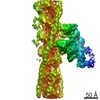

 PDBj
PDBj


 集合体
集合体
 試料調製
試料調製 電子顕微鏡撮影
 FIELD EMISSION GUN / 加速電圧: 300 kV / 照射モード: FLOOD BEAM
 解析
解析