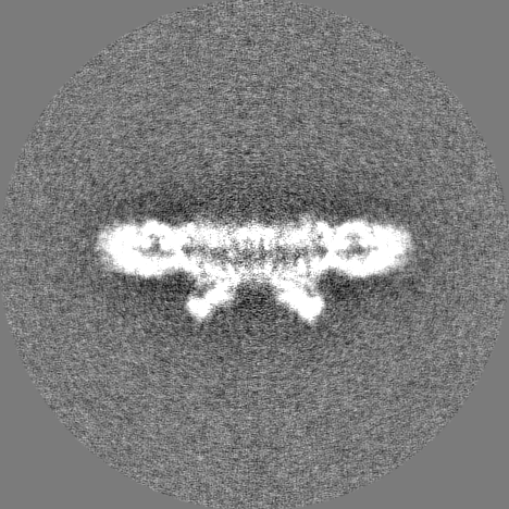
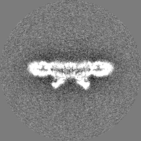
- EMDB-3491: Cryo-EM structure of the PSII supercomplex from Arabidopsis thaliana -
+
Open data
ID or keywords:
Loading...
-
Basic information
Entry
Database: EMDB / ID: EMD-3491
Title
Cryo-EM structure of the PSII supercomplex from Arabidopsis thaliana
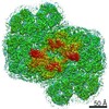
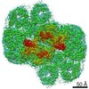
Map data
Sample
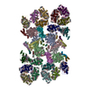
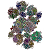
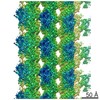
Complex: C2S2M2 supercomplex of Photosystem II
Protein or peptide: x 20 types
Ligand: x 5 types
Keywords
photosynthesis / photosystem II supercomplex / single particle analysis
Function / homology
Function and homology information
chloroplast photosystem II / photoinhibition / photosystem II antenna complex / nonphotochemical quenching / PSII associated light-harvesting complex II / chloroplast stromal thylakoid / thylakoid lumen / plastid thylakoid membrane / plastoglobule / thylakoid membrane ...chloroplast photosystem II / photoinhibition / photosystem II antenna complex / nonphotochemical quenching / PSII associated light-harvesting complex II / chloroplast stromal thylakoid / thylakoid lumen / plastid thylakoid membrane / plastoglobule / thylakoid membrane / chloroplast thylakoid / photosynthesis, light harvesting in photosystem I / photosystem II oxygen evolving complex / photosystem II assembly / apoplast / chloroplast thylakoid lumen / thylakoid / oxygen evolving activity / photosystem II stabilization / protein phosphatase regulator activity / photosystem II reaction center / photosystem II / chloroplast envelope / photosynthetic electron transport chain / oxidoreductase activity, acting on diphenols and related substances as donors, oxygen as acceptor / photosystem I / response to herbicide / photosystem II / poly(U) RNA binding / chloroplast stroma / plastid / photosynthetic electron transport in photosystem II / chlorophyll binding / photosynthesis, light reaction / phosphate ion binding / chloroplast thylakoid membrane / response to light stimulus / photosynthesis / chloroplast / electron transfer activity / protein stabilization / iron ion binding / protein domain specific binding / mRNA binding / heme binding / metal ion binding / nucleus / plasma membrane / cytosol Similarity search - Function
Photosystem II PsbW, class 2 / Photosystem II reaction centre W protein (PsbW) / Photosystem II PsbO, manganese-stabilising / Manganese-stabilising protein / photosystem II polypeptide / Photosystem II reaction centre M protein (PsbM) / Photosystem II PsbM superfamily / Photosystem II PsbM / Photosystem II PsbZ, reaction centre / Photosystem II PsbZ superfamily / YCF9 ...Photosystem II PsbW, class 2 / Photosystem II reaction centre W protein (PsbW) / Photosystem II PsbO, manganese-stabilising / Manganese-stabilising protein / photosystem II polypeptide / Photosystem II reaction centre M protein (PsbM) / Photosystem II PsbM superfamily / Photosystem II PsbM / Photosystem II PsbZ, reaction centre / Photosystem II PsbZ superfamily / YCF9 / Photosystem II PsbX / Photosystem II reaction centre X protein (PsbX) / Photosystem II PsbT / Photosystem II PsbL / Photosystem II CP43 reaction centre protein / Photosystem II PsbL superfamily / Photosystem II PsbT superfamily / Photosystem II CP43 reaction centre protein superfamily / Photosystem II reaction centre T protein / PsbL protein / Photosystem II PsbK / Photosystem II PsbK superfamily / Photosystem II 4 kDa reaction centre component / Photosystem II PsbI / Photosystem II CP47 reaction centre protein / Photosystem II PsbI superfamily / Photosystem II reaction centre I protein (PSII 4.8 kDa protein) / Photosystem II protein D1 / Photosystem II reaction centre protein H / Photosystem II D2 protein / Photosystem II cytochrome b559, conserved site / Photosystem II cytochrome b559, alpha subunit / Photosystem II cytochrome b559, beta subunit / Photosystem II cytochrome b559, N-terminal / Photosystem II cytochrome b559, alpha subunit, lumenal region / Photosystem II reaction centre protein H superfamily / Photosystem II cytochrome b559, alpha subunit superfamily / Cytochrome b559, alpha (gene psbE) and beta (gene psbF)subunits / Lumenal portion of Cytochrome b559, alpha (gene psbE) subunit / Photosystem II 10 kDa phosphoprotein / Cytochrome b559 subunits heme-binding site signature. / : / Photosystem antenna protein-like / Photosystem antenna protein-like superfamily / Photosystem II protein / Chlorophyll A-B binding protein, plant and chromista / Chlorophyll A-B binding protein / Chlorophyll A-B binding protein / Outer membrane protein/outer membrane enzyme PagP, beta-barrel / : / Photosynthetic reaction centre, L/M / Photosystem II protein D1/D2 superfamily / Photosynthetic reaction centre protein / Photosynthetic reaction center proteins signature. Similarity search - Domain/homology
Chlorophyll a-b binding protein 1, chloroplastic / Oxygen-evolving enhancer protein 1-1, chloroplastic / Photosystem II D2 protein / Photosystem II CP47 reaction center protein / Photosystem II CP43 reaction center protein / Cytochrome b559 subunit alpha / Photosystem II reaction center protein H / Photosystem II reaction center protein K / Photosystem II reaction center protein Z / Photosystem II reaction center protein L ...Chlorophyll a-b binding protein 1, chloroplastic / Oxygen-evolving enhancer protein 1-1, chloroplastic / Photosystem II D2 protein / Photosystem II CP47 reaction center protein / Photosystem II CP43 reaction center protein / Cytochrome b559 subunit alpha / Photosystem II reaction center protein H / Photosystem II reaction center protein K / Photosystem II reaction center protein Z / Photosystem II reaction center protein L / Photosystem II reaction center protein T / Cytochrome b559 subunit beta / Photosystem II reaction center protein I / Photosystem II reaction center protein M / Photosystem II protein D1 / Chlorophyll a-b binding protein CP29.1, chloroplastic / Photosystem II reaction center W protein, chloroplastic / Chlorophyll a-b binding protein, chloroplastic / Expressed protein / Chlorophyll a-b binding protein CP26, chloroplastic Similarity search - Component
Biological species
Arabidopsis thaliana (thale cress)
Method
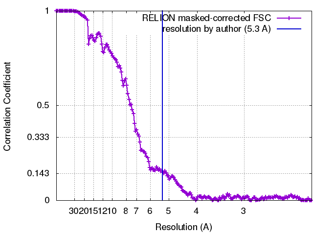
single particle reconstruction / cryo EM / Resolution: 5.3 Å
Journal: Nat Plants / Year: 2017 Title: Subunit and chlorophyll organization of the plant photosystem II supercomplex. Authors: Laura S van Bezouwen / Stefano Caffarri / Ravindra S Kale / Roman Kouřil / Andy-Mark W H Thunnissen / Gert T Oostergetel / Egbert J Boekema / Abstract: Photosystem II (PSII) is a light-driven protein, involved in the primary reactions of photosynthesis. In plant photosynthetic membranes PSII forms large multisubunit supercomplexes, containing a ...Photosystem II (PSII) is a light-driven protein, involved in the primary reactions of photosynthesis. In plant photosynthetic membranes PSII forms large multisubunit supercomplexes, containing a dimeric core and up to four light-harvesting complexes (LHCs), which act as antenna proteins. Here we solved a three-dimensional (3D) structure of the CSM supercomplex from Arabidopsis thaliana using cryo-transmission electron microscopy (cryo-EM) and single-particle analysis at an overall resolution of 5.3 Å. Using a combination of homology modelling and restrained refinement against the cryo-EM map, it was possible to model atomic structures for all antenna complexes and almost all core subunits. We located all 35 chlorophylls of the core region based on the cyanobacterial PSII structure, whose positioning is highly conserved, as well as all the chlorophylls of the LHCII S and M trimers. A total of 13 and 9 chlorophylls were identified in CP26 and CP24, respectively. Energy flow from LHC complexes to the PSII reaction centre is proposed to follow preferential pathways: CP26 and CP29 directly transfer to the core using several routes for efficient transfer; the S trimer is directly connected to CP43 and the M trimer can efficiently transfer energy to the core through CP29 and the S trimer.
History
Deposition
Nov 13, 2016
-
Header (metadata) release
Jun 21, 2017
-
Map release
Jun 21, 2017
-
Update
Nov 20, 2024
-
Current status
Nov 20, 2024
Processing site: PDBe / Status: Released
-
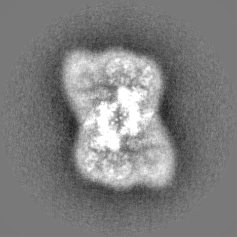
Structure visualization
Movie
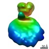
Surface view with section colored by density value
Cryogen name: ETHANE / Chamber humidity: 100 % / Chamber temperature: 293 K / Instrument: FEI VITROBOT MARK III
-
Electron microscopy
Microscope
FEI TITAN KRIOS
Specialist optics
Spherical aberration corrector: Microscope had a Cs corrector
Image recording
Film or detector model: FEI FALCON II (4k x 4k) / Detector mode: INTEGRATING / Digitization - Frames/image: 1-7 / Number grids imaged: 3 / Number real images: 5198 / Average exposure time: 1.0 sec. / Average electron dose: 38.0 e/Å2
Electron beam
Acceleration voltage: 300 kV / Electron source: FIELD EMISSION GUN
Initial fitting of the subunits in the cryo-EM map was performed by rigid body real space refinement, using as templates the high resolution crystal structures of Thermosynechococcus vulcanus PSII (PDB code 3WU2), pea LHC-II (PDB code 2BHW for the S- and M-trimers, and spinach CP29 (PDB code 3PL9) for CP29, CP26 and CP24. Local fitting and adjustment of the subunits in the cryo-EM maps was performed using manual rebuilding and restrained real space refinement as explained in the primary reference. Due to large differences in local resolution of the cryo-EM map, refinement of the PSII core, the S-trimer with CP26/CP29 and the M-trimer with CP24 was performed separately in excised parts of the cryo-EM map. The core was refined at 4.5 angstrom, the S-trimer+CP26+CP29 at 5.5 angstrom and the M-trimer+CP24 at 6.5 angstrom. Core: chains A,B,C,D,E,F,H,I,J,K,L,M,O,T,W,X,Z and a,b,c,d,e,f,h,i,j,k,l,m,o,t,w,x,z. S-trimer+CP26+CP29: chains G,N,Y,S,R and g,n,y,s,r. M-trimer+CP24: chains 1,2,3,4 and 5,6,7,8.
Refinement
Space: REAL / Protocol: FLEXIBLE FIT / Target criteria: Cross-correlation coefficient
Output model
PDB-5mdx: Cryo-EM structure of the PSII supercomplex from Arabidopsis thaliana
+
About Yorodumi
-
News
-
Feb 9, 2022. New format data for meta-information of EMDB entries
New format data for meta-information of EMDB entries
Version 3 of the EMDB header file is now the official format.
The previous official version 1.9 will be removed from the archive.
In the structure databanks used in Yorodumi, some data are registered as the other names, "COVID-19 virus" and "2019-nCoV". Here are the details of the virus and the list of structure data.
Jan 31, 2019. EMDB accession codes are about to change! (news from PDBe EMDB page)
EMDB accession codes are about to change! (news from PDBe EMDB page)
The allocation of 4 digits for EMDB accession codes will soon come to an end. Whilst these codes will remain in use, new EMDB accession codes will include an additional digit and will expand incrementally as the available range of codes is exhausted. The current 4-digit format prefixed with “EMD-” (i.e. EMD-XXXX) will advance to a 5-digit format (i.e. EMD-XXXXX), and so on. It is currently estimated that the 4-digit codes will be depleted around Spring 2019, at which point the 5-digit format will come into force.
The EM Navigator/Yorodumi systems omit the EMD- prefix.
Related info.:Q: What is EMD? / ID/Accession-code notation in Yorodumi/EM Navigator
Yorodumi is a browser for structure data from EMDB, PDB, SASBDB, etc.
This page is also the successor to EM Navigator detail page, and also detail information page/front-end page for Omokage search.
The word "yorodu" (or yorozu) is an old Japanese word meaning "ten thousand". "mi" (miru) is to see.
Related info.:EMDB / PDB / SASBDB / Comparison of 3 databanks / Yorodumi Search / Aug 31, 2016. New EM Navigator & Yorodumi / Yorodumi Papers / Jmol/JSmol / Function and homology information / Changes in new EM Navigator and Yorodumi
 Movie
Movie Controller

 Yorodumi
 Open data
 Basic information
 Map data
 Sample
 Keywords
 Function and homology information
 Authors
 Citation


 Structure visualization
 Downloads & links
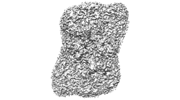 emd_3491.png
 http://ftp.pdbj.org/pub/emdb/structures/EMD-3491


 Z (Sec.)
 Y (Row.)
 X (Col.)


 Sample components


 Processing
 Electron microscopy
 FIELD EMISSION GUN