Movie
Movie Controller

[English] 日本語
 Yorodumi
- PDB-7n65: Complex structure of HIV superinfection Fab QA013.2 and BG505.SOS... -
+ Open data

- Basic information
| Entry | Database: PDB / ID: 7n65 | ||||||
|---|---|---|---|---|---|---|---|
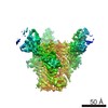
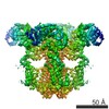
| Title | Complex structure of HIV superinfection Fab QA013.2 and BG505.SOSIP.664 | ||||||
 Components Components |
| ||||||
 Keywords Keywords | VIRAL PROTEIN/IMMUNE SYSTEM / HIV-1 Envelope / superinfection antibody QA013.2 / VIRAL PROTEIN / VIRAL PROTEIN-IMMUNE SYSTEM complex | ||||||
| Function / homology |  Function and homology information Function and homology informationmembrane fusion involved in viral entry into host cell / symbiont-mediated perturbation of host defense response / positive regulation of plasma membrane raft polarization / positive regulation of receptor clustering / host cell endosome membrane / clathrin-dependent endocytosis of virus by host cell / viral protein processing / fusion of virus membrane with host plasma membrane / fusion of virus membrane with host endosome membrane / viral envelope ...membrane fusion involved in viral entry into host cell / symbiont-mediated perturbation of host defense response / positive regulation of plasma membrane raft polarization / positive regulation of receptor clustering / host cell endosome membrane / clathrin-dependent endocytosis of virus by host cell / viral protein processing / fusion of virus membrane with host plasma membrane / fusion of virus membrane with host endosome membrane / viral envelope / symbiont entry into host cell / virion attachment to host cell / host cell plasma membrane / virion membrane / structural molecule activity / membrane Similarity search - Function | ||||||
| Biological species |   Human immunodeficiency virus 1 Human immunodeficiency virus 1 Homo sapiens (human) Homo sapiens (human) | ||||||
| Method | ELECTRON MICROSCOPY / single particle reconstruction / cryo EM / Resolution: 4.15 Å | ||||||
 Authors Authors | Mangala Prasad, V. / Shipley, M.M. / Overbaugh, J.M. / Lee, K.K. | ||||||
| Funding support |  United States, 1items United States, 1items
| ||||||
 Citation Citation | Journal: Elife / Year: 2021 Title: Functional development of a V3/glycan-specific broadly neutralizing antibody isolated from a case of HIV superinfection. Authors: Mackenzie M Shipley / Vidya Mangala Prasad / Laura E Doepker / Adam Dingens / Duncan K Ralph / Elias Harkins / Amrit Dhar / Dana Arenz / Vrasha Chohan / Haidyn Weight / Kishor Mandaliya / ...Authors: Mackenzie M Shipley / Vidya Mangala Prasad / Laura E Doepker / Adam Dingens / Duncan K Ralph / Elias Harkins / Amrit Dhar / Dana Arenz / Vrasha Chohan / Haidyn Weight / Kishor Mandaliya / Jesse D Bloom / Frederick A Matsen / Kelly K Lee / Julie M Overbaugh /  Abstract: Stimulating broadly neutralizing antibodies (bnAbs) directly from germline remains a barrier for HIV vaccines. HIV superinfection elicits bnAbs more frequently than single infection, providing clues ...Stimulating broadly neutralizing antibodies (bnAbs) directly from germline remains a barrier for HIV vaccines. HIV superinfection elicits bnAbs more frequently than single infection, providing clues of how to elicit such responses. We used longitudinal antibody sequencing and structural studies to characterize bnAb development from a superinfection case. BnAb QA013.2 bound initial and superinfecting viral Env, despite its probable naive progenitor only recognizing the superinfecting strain, suggesting both viruses influenced this lineage. A 4.15 Å cryo-EM structure of QA013.2 bound to native-like trimer showed recognition of V3 signatures (N301/N332 and GDIR). QA013.2 relies less on CDRH3 and more on framework and CDRH1 for affinity and breadth compared to other V3/glycan-specific bnAbs. Antigenic profiling revealed that viral escape was achieved by changes in the structurally-defined epitope and by mutations in V1. These results highlight shared and novel properties of QA013.2 relative to other V3/glycan-specific bnAbs in the setting of sequential, diverse antigens. | ||||||
| History |
|
- Structure visualization
| Movie |
Movie viewer |
|---|---|
| Structure viewer | Molecule: MolmilJmol/JSmol |
- Downloads & links
-Download
| PDBx/mmCIF format | 7n65.cif.gz | 494 KB | Display | PDBx/mmCIF format |
|---|---|---|---|---|
| PDB format | pdb7n65.ent.gz | Display | PDB format | |
| PDBx/mmJSON format | 7n65.json.gz | Tree view | PDBx/mmJSON format | |
| Others |  Other downloads Other downloads |
-Validation report
| Arichive directory | https://data.pdbj.org/pub/pdb/validation_reports/n6/7n65ftp://data.pdbj.org/pub/pdb/validation_reports/n6/7n65 | HTTPS FTP |
|---|
-Related structure data
| Related structure data |  24195MC M: map data used to model this data C: citing same article ( |
|---|---|
| Similar structure data |
-Links
 PDBj
PDBj


- Assembly
| Deposited unit | 
|
|---|---|
| 1 |
|
-Components
-Envelope glycoprotein ... , 2 types, 6 molecules AEIBFJ
| #1: Protein | Mass: 56846.613 Da / Num. of mol.: 3 / Mutation: T332N Source method: isolated from a genetically manipulated source Source: (gene. exp.) Human immunodeficiency virus 1 / Strain: BG505 / Gene: env / Cell line (production host): Expi293F / Production host: Homo sapiens (human) / References: UniProt: A0A6H1VCM1#2: Protein | Mass: 18245.830 Da / Num. of mol.: 3 Source method: isolated from a genetically manipulated source Source: (gene. exp.) Human immunodeficiency virus 1 / Strain: BG505 / Gene: env / Cell line (production host): Expi 293F / Production host: Homo sapiens (human) / References: UniProt: Q2N0S7 |
|---|
-Antibody , 2 types, 6 molecules CGKDHL
| #3: Antibody | Mass: 14384.085 Da / Num. of mol.: 3 Source method: isolated from a genetically manipulated source Source: (gene. exp.) Homo sapiens (human) / Cell line: Peripheral blood mononuclear cell / Cell line (production host): HEK 293F / Production host: Homo sapiens (human)#4: Antibody | Mass: 11189.263 Da / Num. of mol.: 3 Source method: isolated from a genetically manipulated source Source: (gene. exp.) Homo sapiens (human) / Cell line: Peripheral blood mononuclear cell / Cell line (production host): HEK 293F / Production host: Homo sapiens (human) |
|---|
-Sugars , 8 types, 63 molecules 

| #5: Polysaccharide | beta-D-mannopyranose-(1-4)-2-acetamido-2-deoxy-beta-D-glucopyranose-(1-4)-2-acetamido-2-deoxy-beta- ...beta-D-mannopyranose-(1-4)-2-acetamido-2-deoxy-beta-D-glucopyranose-(1-4)-2-acetamido-2-deoxy-beta-D-glucopyranose Source method: isolated from a genetically manipulated source #6: Polysaccharide | 2-acetamido-2-deoxy-beta-D-glucopyranose-(1-4)-2-acetamido-2-deoxy-beta-D-glucopyranose Source method: isolated from a genetically manipulated source #7: Polysaccharide | Source method: isolated from a genetically manipulated source #8: Polysaccharide | Source method: isolated from a genetically manipulated source #9: Polysaccharide | Source method: isolated from a genetically manipulated source #10: Polysaccharide | Source method: isolated from a genetically manipulated source #11: Sugar | ChemComp-NAG /  Type: D-saccharide, beta linking / Mass: 221.208 Da / Num. of mol.: 9 Type: D-saccharide, beta linking / Mass: 221.208 Da / Num. of mol.: 9Source method: isolated from a genetically manipulated source Formula: C8H15NO6 #12: Sugar |  Type: D-saccharide, alpha linking / Mass: 180.156 Da / Num. of mol.: 3 / Source method: obtained synthetically / Formula: C6H12O6 Type: D-saccharide, alpha linking / Mass: 180.156 Da / Num. of mol.: 3 / Source method: obtained synthetically / Formula: C6H12O6 |
|---|
-Details
| Has ligand of interest | N |
|---|---|
| Has protein modification | Y |
-Experimental details
-Experiment
| Experiment | Method: ELECTRON MICROSCOPY |
|---|---|
| EM experiment | Aggregation state: PARTICLE / 3D reconstruction method: single particle reconstruction |
- Sample preparation
| Component | Name: Complex of HIV-1 envelope protein with super-infection Fab QA013.2 Type: COMPLEX / Entity ID: #1-#4 / Source: RECOMBINANT | |||||||||||||||
|---|---|---|---|---|---|---|---|---|---|---|---|---|---|---|---|---|
| Molecular weight | Value: 0.5 MDa / Experimental value: NO | |||||||||||||||
| Source (natural) |
| |||||||||||||||
| Source (recombinant) | Organism: Homo sapiens (human) / Cell: 293F | |||||||||||||||
| Buffer solution | pH: 7.4 / Details: 1X PBS at pH 7.4, made fresh. | |||||||||||||||
| Buffer component | Name: Phosphate buffer saline | |||||||||||||||
| Specimen | Conc.: 0.05 mg/ml / Embedding applied: NO / Shadowing applied: NO / Staining applied: NO / Vitrification applied: YES / Details: Monodisperse sample in PBS | |||||||||||||||
| Specimen support | Grid material: COPPER / Grid mesh size: 400 divisions/in. / Grid type: PELCO Ultrathin Carbon with Lacey Carbon | |||||||||||||||
| Vitrification | Instrument: FEI VITROBOT MARK IV / Cryogen name: ETHANE / Humidity: 100 % / Chamber temperature: 277 K / Details: 3ul sample volume, blotted for 3-4 seconds |
- Electron microscopy imaging
| Experimental equipment |  Model: Titan Krios / Image courtesy: FEI Company |
|---|---|
| Microscopy | Model: FEI TITAN KRIOS / Details: Preliminary grid screening was performed manually |
| Electron gun | Electron source:  FIELD EMISSION GUN / Accelerating voltage: 300 kV / Illumination mode: FLOOD BEAM FIELD EMISSION GUN / Accelerating voltage: 300 kV / Illumination mode: FLOOD BEAM |
| Electron lens | Mode: BRIGHT FIELD / Nominal magnification: 110000 X / Nominal defocus max: 3500 nm / Nominal defocus min: 1700 nm / Cs: 2.7 mm / C2 aperture diameter: 50 µm / Alignment procedure: COMA FREE |
| Specimen holder | Cryogen: NITROGEN / Specimen holder model: FEI TITAN KRIOS AUTOGRID HOLDER |
| Image recording | Average exposure time: 10 sec. / Electron dose: 44 e/Å2 / Detector mode: COUNTING / Film or detector model: GATAN K2 SUMMIT (4k x 4k) / Num. of real images: 6475 |
| Image scans | Movie frames/image: 50 |
- Processing
| Software | Name: PHENIX / Version: 1.17.1_3660: / Classification: refinement | ||||||||||||||||||||||||||||
|---|---|---|---|---|---|---|---|---|---|---|---|---|---|---|---|---|---|---|---|---|---|---|---|---|---|---|---|---|---|
| EM software |
| ||||||||||||||||||||||||||||
| CTF correction | Type: PHASE FLIPPING AND AMPLITUDE CORRECTION | ||||||||||||||||||||||||||||
| Particle selection | Num. of particles selected: 1082742 / Details: LOG picker | ||||||||||||||||||||||||||||
| Symmetry | Point symmetry: C3 (3 fold cyclic) | ||||||||||||||||||||||||||||
| 3D reconstruction | Resolution: 4.15 Å / Resolution method: FSC 0.143 CUT-OFF / Num. of particles: 113470 / Algorithm: FOURIER SPACE / Symmetry type: POINT | ||||||||||||||||||||||||||||
| Atomic model building | Protocol: RIGID BODY FIT Details: Rigid body fitting in Chimera, followed by real space refinement in Phenix using reference model restraints | ||||||||||||||||||||||||||||
| Atomic model building | PDB-ID: 5ACO Accession code: 5ACO / Source name: PDB / Type: experimental model | ||||||||||||||||||||||||||||
| Refinement | Cross valid method: NONE Stereochemistry target values: GeoStd + Monomer Library + CDL v1.2 | ||||||||||||||||||||||||||||
| Displacement parameters | Biso mean: 124.29 Å2 | ||||||||||||||||||||||||||||
| Refine LS restraints |
|