 Movie
Movie Controller

[English] 日本語
 Yorodumi
- PDB-5aco: Cryo-EM structure of PGT128 Fab in complex with BG505 SOSIP.664 E... -
+ Open data

- Basic information
| Entry | Database: PDB / ID: 5aco | |||||||||
|---|---|---|---|---|---|---|---|---|---|---|
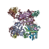
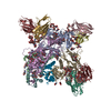
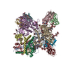
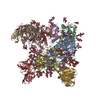
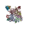
| Title | Cryo-EM structure of PGT128 Fab in complex with BG505 SOSIP.664 Env trimer | |||||||||
 Components Components |
| |||||||||
 Keywords Keywords | VIRAL PROTEIN / IMMUNE SYSTEM / HIV-1 / ENV / BNAB / ANTIBODY / PGT128 | |||||||||
| Function / homology |  Function and homology information Function and homology informationsymbiont-mediated perturbation of host defense response / positive regulation of plasma membrane raft polarization / positive regulation of receptor clustering / host cell endosome membrane / clathrin-dependent endocytosis of virus by host cell / viral protein processing / fusion of virus membrane with host plasma membrane / fusion of virus membrane with host endosome membrane / viral envelope / virion attachment to host cell ...symbiont-mediated perturbation of host defense response / positive regulation of plasma membrane raft polarization / positive regulation of receptor clustering / host cell endosome membrane / clathrin-dependent endocytosis of virus by host cell / viral protein processing / fusion of virus membrane with host plasma membrane / fusion of virus membrane with host endosome membrane / viral envelope / virion attachment to host cell / host cell plasma membrane / virion membrane / structural molecule activity / membrane / identical protein binding Similarity search - Function | |||||||||
| Biological species |   HUMAN IMMUNODEFICIENCY VIRUS 1 HUMAN IMMUNODEFICIENCY VIRUS 1 HOMO SAPIENS (human) HOMO SAPIENS (human) | |||||||||
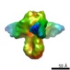
| Method | ELECTRON MICROSCOPY / single particle reconstruction / cryo EM / Resolution: 4.36 Å | |||||||||
 Authors Authors | Lee, J.H. / Ward, A.B. | |||||||||
 Citation Citation | Journal: Structure / Year: 2015 Title: Model Building and Refinement of a Natively Glycosylated HIV-1 Env Protein by High-Resolution Cryoelectron Microscopy. Authors: Jeong Hyun Lee / Natalia de Val / Dmitry Lyumkis / Andrew B Ward /  Abstract: Secretory and membrane proteins from mammalian cells undergo post-translational modifications, including N-linked glycosylation, which can result in a large number of possible glycoforms. This sample ...Secretory and membrane proteins from mammalian cells undergo post-translational modifications, including N-linked glycosylation, which can result in a large number of possible glycoforms. This sample heterogeneity can be problematic for structural studies, particularly X-ray crystallography. Thus, crystal structures of heavily glycosylated proteins such as the HIV-1 Env viral spike protein have been determined by removing the majority of glycans. This step is most frequently carried out using Endoglycosidase H (EndoH) and requires that all expressed glycans be in the high-mannose form, which is often not the native glycoform. With significantly improved technologies in single-particle cryoelectron microscopy, we demonstrate that it is now possible to refine and build natively glycosylated HIV-1 Env structures in solution to 4.36 Å resolution. At this resolution we can now analyze the complete epitope of a broadly neutralizing antibody (bnAb), PGT128, in the context of the trimer expressed with native glycans. | |||||||||
| History |
|
- Structure visualization
| Movie |
Movie viewer |
|---|---|
| Structure viewer | Molecule: MolmilJmol/JSmol |
- Downloads & links
-Download
| PDBx/mmCIF format | 5aco.cif.gz | 479.2 KB | Display | PDBx/mmCIF format |
|---|---|---|---|---|
| PDB format | pdb5aco.ent.gz | Display | PDB format | |
| PDBx/mmJSON format | 5aco.json.gz | Tree view | PDBx/mmJSON format | |
| Others |  Other downloads Other downloads |
-Validation report
| Arichive directory | https://data.pdbj.org/pub/pdb/validation_reports/ac/5acoftp://data.pdbj.org/pub/pdb/validation_reports/ac/5aco | HTTPS FTP |
|---|
-Related structure data
| Related structure data | 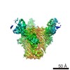 3121MC 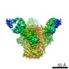 3120C M: map data used to model this data C: citing same article ( |
|---|---|
| Similar structure data |

-Links
 PDBj
PDBj


- Assembly
| Deposited unit | 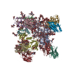
|
|---|---|
| 1 |
|
-Components
-HIV-1 ENVELOPE ... , 2 types, 6 molecules ACDBEF
| #1: Protein | Mass: 53278.301 Da / Num. of mol.: 3 / Fragment: GP120, RESIDUES 30-505 / Mutation: YES Source method: isolated from a genetically manipulated source Details: THE ENV SEQUENCE IS FROM THE CLADE A VIRUS BG505, TRUNCATED AT RESIDUE 664 OF GP41, MUTATED TO HAVE THE N332 GLYCOSYLATION SITE, AND CONTAINS STABILIZING SOSIP MUTATIONS. Source: (gene. exp.) HUMAN IMMUNODEFICIENCY VIRUS 1 / Gene: ENV / Variant: BG505 SOSIP.664 / Cell line (production host): HEK293 / Production host: HOMO SAPIENS (human) / References: UniProt: Q2N0S6#2: Protein | Mass: 17146.482 Da / Num. of mol.: 3 / Fragment: GP41, RESIDUES 509-661 / Mutation: YES Source method: isolated from a genetically manipulated source Details: THE ENV SEQUENCE IS FROM THE CLADE A VIRUS BG505, TRUNCATED AT RESIDUE 664 OF GP41, MUTATED TO HAVE THE N332 GLYCOSYLATION SITE, AND CONTAINS STABILIZING SOSIP MUTATIONS. Source: (gene. exp.) HUMAN IMMUNODEFICIENCY VIRUS 1 / Gene: ENV / Variant: BG505 SOSIP.664 / Cell line (production host): HEK293 / Production host: HOMO SAPIENS (human) / References: UniProt: Q2N0S6 |
|---|
-Antibody , 2 types, 6 molecules GHIJKL
| #3: Antibody | Mass: 25580.701 Da / Num. of mol.: 3 / Fragment: HEAVY CHAIN OF FAB VARIABLE REGION Source method: isolated from a genetically manipulated source Details: THE FRAGMENT ANTIGEN BINDING (FAB) OF BNAB PGT128. / Source: (gene. exp.) HOMO SAPIENS (human) / Cell line (production host): HEK293 / Production host: HOMO SAPIENS (human)#4: Antibody | Mass: 22224.572 Da / Num. of mol.: 3 / Fragment: LIGHT CHAIN OF FAB VARIABLE REGION Source method: isolated from a genetically manipulated source Details: THE FRAGMENT ANTIGEN BINDING (FAB) OF BNAB PGT128. / Source: (gene. exp.) HOMO SAPIENS (human) / Cell line (production host): HEK293 / Production host: HOMO SAPIENS (human) |
|---|
-Sugars , 6 types, 60 molecules 
| #5: Polysaccharide | beta-D-mannopyranose-(1-4)-2-acetamido-2-deoxy-beta-D-glucopyranose-(1-4)-2-acetamido-2-deoxy-beta- ...beta-D-mannopyranose-(1-4)-2-acetamido-2-deoxy-beta-D-glucopyranose-(1-4)-2-acetamido-2-deoxy-beta-D-glucopyranose Source method: isolated from a genetically manipulated source #6: Polysaccharide | 2-acetamido-2-deoxy-beta-D-glucopyranose-(1-4)-2-acetamido-2-deoxy-beta-D-glucopyranose Source method: isolated from a genetically manipulated source #7: Polysaccharide | alpha-D-mannopyranose-(1-2)-alpha-D-mannopyranose-(1-3)-[alpha-D-mannopyranose-(1-3)-alpha-D- ...alpha-D-mannopyranose-(1-2)-alpha-D-mannopyranose-(1-3)-[alpha-D-mannopyranose-(1-3)-alpha-D-mannopyranose-(1-6)]beta-D-mannopyranose-(1-4)-2-acetamido-2-deoxy-beta-D-glucopyranose-(1-4)-2-acetamido-2-deoxy-beta-D-glucopyranose Source method: isolated from a genetically manipulated source #8: Polysaccharide | Source method: isolated from a genetically manipulated source #9: Polysaccharide | Source method: isolated from a genetically manipulated source #10: Sugar | ChemComp-NAG /  Type: D-saccharide, beta linking / Mass: 221.208 Da / Num. of mol.: 9 Type: D-saccharide, beta linking / Mass: 221.208 Da / Num. of mol.: 9Source method: isolated from a genetically manipulated source Formula: C8H15NO6 |
|---|
-Details
| Has protein modification | Y |
|---|
-Experimental details
-Experiment
| Experiment | Method: ELECTRON MICROSCOPY |
|---|---|
| EM experiment | Aggregation state: PARTICLE / 3D reconstruction method: single particle reconstruction |
- Sample preparation
| Component | Name: PGT128 FAB BOUND TO BG505 SOSIP.664 ENV TRIMER / Type: COMPLEX |
|---|---|
| Buffer solution | Name: 50 MM TRIS, 150 MM NACL, 0.675 MM DDM / pH: 7.4 / Details: 50 MM TRIS, 150 MM NACL, 0.675 MM DDM |
| Specimen | Conc.: 2.5 mg/ml / Embedding applied: NO / Shadowing applied: NO / Staining applied: NO / Vitrification applied: YES |
| Specimen support | Details: HOLEY CARBON |
| Vitrification | Instrument: HOMEMADE PLUNGER / Cryogen name: ETHANE / Details: FROZEN IN LIQUID ETHANE AT 4 DEGREES C. |
- Electron microscopy imaging
| Experimental equipment |  Model: Titan Krios / Image courtesy: FEI Company |
|---|---|
| Microscopy | Model: FEI TITAN KRIOS / Date: Oct 7, 2014 / Details: IMAGED ON FEI TITAN KRIOS |
| Electron gun | Electron source:  FIELD EMISSION GUN / Accelerating voltage: 300 kV / Illumination mode: FLOOD BEAM FIELD EMISSION GUN / Accelerating voltage: 300 kV / Illumination mode: FLOOD BEAM |
| Electron lens | Mode: BRIGHT FIELD / Nominal magnification: 22500 X / Calibrated magnification: 22500 X / Nominal defocus max: 3500 nm / Nominal defocus min: 1000 nm / Cs: 2.7 mm |
| Specimen holder | Tilt angle min: 0 ° |
| Image recording | Electron dose: 35 e/Å2 / Film or detector model: GATAN K2 SUMMIT (4k x 4k) |
| Image scans | Num. digital images: 2000 |
- Processing
| EM software | Name: RELION / Category: 3D reconstruction | ||||||||||||
|---|---|---|---|---|---|---|---|---|---|---|---|---|---|
| CTF correction | Details: WHOLE MICROGRAPH | ||||||||||||
| Symmetry | Point symmetry: C3 (3 fold cyclic) | ||||||||||||
| 3D reconstruction | Method: MAXIMUM LIKELIHOOD / Resolution: 4.36 Å / Num. of particles: 92095 / Nominal pixel size: 1.31 Å / Actual pixel size: 1.31 Å Details: SUBMISSION BASED ON EXPERIMENTAL DATA FROM EMDB EMD-3121. (DEPOSITION ID: 13671). Symmetry type: POINT | ||||||||||||
| Atomic model building | Protocol: OTHER / Space: REAL / Details: METHOD--GLOBAL REFINEMENT PROTOCOL--CRYOEM | ||||||||||||
| Refinement | Highest resolution: 4.36 Å | ||||||||||||
| Refinement step | Cycle: LAST / Highest resolution: 4.36 Å
|
