 Movie
Movie Controller

[English] 日本語
 Yorodumi
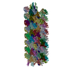
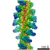
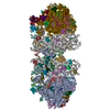
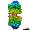
- PDB-7moq: The structure of the Tetrahymena thermophila outer dynein arm on ... -
+ Open data

- Basic information
| Entry | Database: PDB / ID: 7moq | |||||||||
|---|---|---|---|---|---|---|---|---|---|---|
| Title | The structure of the Tetrahymena thermophila outer dynein arm on doublet microtubule | |||||||||
 Components Components |
| |||||||||
 Keywords Keywords | STRUCTURAL PROTEIN / cilia / doublet / axoneme / outer dynein arm / dynein | |||||||||
| Function / homology |  Function and homology information Function and homology informationinner dynein arm / axonemal dynein complex / outer dynein arm / outer dynein arm assembly / inner dynein arm assembly / cilium movement involved in cell motility / cilium movement / dynein light chain binding / dynein heavy chain binding / dynein complex ...inner dynein arm / axonemal dynein complex / outer dynein arm / outer dynein arm assembly / inner dynein arm assembly / cilium movement involved in cell motility / cilium movement / dynein light chain binding / dynein heavy chain binding / dynein complex / ciliary rootlet / minus-end-directed microtubule motor activity / dynein light intermediate chain binding / cytoplasmic dynein complex / microtubule-based movement / dynein intermediate chain binding / protein-disulfide reductase activity / axoneme / mRNA transport / microtubule-based process / cell redox homeostasis / ciliary tip / structural constituent of cytoskeleton / protein transport / microtubule binding / microtubule / Hydrolases; Acting on acid anhydrides; Acting on GTP to facilitate cellular and subcellular movement / ciliary basal body / hydrolase activity / GTPase activity / GTP binding / ATP binding / metal ion binding / nucleus / cytoplasm Similarity search - Function | |||||||||
| Biological species |  Tetrahymena thermophila CU428 (eukaryote) Tetrahymena thermophila CU428 (eukaryote) | |||||||||
| Method | ELECTRON MICROSCOPY / single particle reconstruction / cryo EM / Resolution: 8 Å | |||||||||
 Authors Authors | Kubo, S. / Yang, S.K. / Ichikawa, M. / Bui, K.H. | |||||||||
| Funding support |  Canada, 2items Canada, 2items
| |||||||||
 Citation Citation | Journal: EMBO Rep / Year: 2021 Title: Remodeling and activation mechanisms of outer arm dyneins revealed by cryo-EM. Authors: Shintaroh Kubo / Shun Kai Yang / Corbin S Black / Daniel Dai / Melissa Valente-Paterno / Jacek Gaertig / Muneyoshi Ichikawa / Khanh Huy Bui /   Abstract: Cilia are thin microtubule-based protrusions of eukaryotic cells. The swimming of ciliated protists and sperm cells is propelled by the beating of cilia. Cilia propagate the flow of mucus in the ...Cilia are thin microtubule-based protrusions of eukaryotic cells. The swimming of ciliated protists and sperm cells is propelled by the beating of cilia. Cilia propagate the flow of mucus in the trachea and protect the human body from viral infections. The main force generators of ciliary beating are the outer dynein arms (ODAs) which attach to the doublet microtubules. The bending of cilia is driven by the ODAs' conformational changes caused by ATP hydrolysis. Here, we report the native ODA complex structure attaching to the doublet microtubule by cryo-electron microscopy. The structure reveals how the ODA complex is attached to the doublet microtubule via the docking complex in its native state. Combined with coarse-grained molecular dynamic simulations, we present a model of how the attachment of the ODA to the doublet microtubule induces remodeling and activation of the ODA complex. | |||||||||
| History |
|
- Structure visualization
| Movie |
Movie viewer |
|---|---|
| Structure viewer | Molecule: MolmilJmol/JSmol |
- Downloads & links
-Download
| PDBx/mmCIF format | 7moq.cif.gz | 2.9 MB | Display | PDBx/mmCIF format |
|---|---|---|---|---|
| PDB format | pdb7moq.ent.gz | Display | PDB format | |
| PDBx/mmJSON format | 7moq.json.gz | Tree view | PDBx/mmJSON format | |
| Others |  Other downloads Other downloads |
-Validation report
| Arichive directory | https://data.pdbj.org/pub/pdb/validation_reports/mo/7moqftp://data.pdbj.org/pub/pdb/validation_reports/mo/7moq | HTTPS FTP |
|---|
-Related structure data
| Related structure data |  23926MC M: map data used to model this data C: citing same article ( |
|---|---|
| Similar structure data |


-Links
 PDBj
PDBj


- Assembly
| Deposited unit | 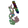
|
|---|---|
| 1 |
|
-Components
-Protein , 7 types, 19 molecules ACDdEePQUYuqyRSWsrw
| #1: Protein | Mass: 475554.406 Da / Num. of mol.: 1 / Source method: isolated from a natural source / Source: (natural) Tetrahymena thermophila CU428 (eukaryote) / Strain: CU428 / References: UniProt: I7M6H4 | ||||||||
|---|---|---|---|---|---|---|---|---|---|
| #3: Protein | Mass: 534328.812 Da / Num. of mol.: 1 / Source method: isolated from a natural source / Source: (natural) Tetrahymena thermophila CU428 (eukaryote) / Strain: CU428 / References: UniProt: Q22A67 | ||||||||
| #4: Protein | Mass: 77888.219 Da / Num. of mol.: 2 / Source method: isolated from a natural source / Source: (natural) Tetrahymena thermophila CU428 (eukaryote) / Strain: CU428 / References: UniProt: I7M008#5: Protein | Mass: 77178.062 Da / Num. of mol.: 2 / Source method: isolated from a natural source / Source: (natural) Tetrahymena thermophila CU428 (eukaryote) / Strain: CU428 / References: UniProt: Q23FU1#16: Protein | | Mass: 12855.699 Da / Num. of mol.: 1 / Source method: isolated from a natural source / Source: (natural) Tetrahymena thermophila CU428 (eukaryote) / Strain: CU428 / References: UniProt: Q1HFX5#17: Protein | Mass: 49617.676 Da / Num. of mol.: 6 / Source method: isolated from a natural source / Source: (natural) Tetrahymena thermophila CU428 (eukaryote) / Strain: CU428 / References: UniProt: P41352#18: Protein | Mass: 49639.035 Da / Num. of mol.: 6 / Source method: isolated from a natural source / Source: (natural) Tetrahymena thermophila CU428 (eukaryote) / Strain: CU428 / References: UniProt: P41351 |
-Dynein light ... , 10 types, 10 molecules FGHIJKLMNO
| #6: Protein | Mass: 14751.817 Da / Num. of mol.: 1 / Source method: isolated from a natural source / Source: (natural) Tetrahymena thermophila CU428 (eukaryote) / Strain: CU428 / References: UniProt: I7MHB1 |
|---|---|
| #7: Protein | Mass: 18490.188 Da / Num. of mol.: 1 / Source method: isolated from a natural source / Source: (natural) Tetrahymena thermophila CU428 (eukaryote) / Strain: CU428 / References: UniProt: Q22MV2 |
| #8: Protein | Mass: 10780.357 Da / Num. of mol.: 1 / Source method: isolated from a natural source / Source: (natural) Tetrahymena thermophila CU428 (eukaryote) / Strain: CU428 / References: UniProt: Q1HFW2 |
| #9: Protein | Mass: 12348.086 Da / Num. of mol.: 1 / Source method: isolated from a natural source / Source: (natural) Tetrahymena thermophila CU428 (eukaryote) / Strain: CU428 / References: UniProt: Q1HFX0 |
| #10: Protein | Mass: 10973.408 Da / Num. of mol.: 1 / Source method: isolated from a natural source / Source: (natural) Tetrahymena thermophila CU428 (eukaryote) / Strain: CU428 / References: UniProt: Q1HFV9 |
| #11: Protein | Mass: 13336.089 Da / Num. of mol.: 1 / Source method: isolated from a natural source / Source: (natural) Tetrahymena thermophila CU428 (eukaryote) / Strain: CU428 / References: UniProt: Q22R86 |
| #12: Protein | Mass: 12516.457 Da / Num. of mol.: 1 / Source method: isolated from a natural source / Source: (natural) Tetrahymena thermophila CU428 (eukaryote) / Strain: CU428 / References: UniProt: W7XJB1 |
| #13: Protein | Mass: 10453.167 Da / Num. of mol.: 1 / Source method: isolated from a natural source / Source: (natural) Tetrahymena thermophila CU428 (eukaryote) / Strain: CU428 / References: UniProt: Q1HFW0 |
| #14: Protein | Mass: 15608.120 Da / Num. of mol.: 1 / Source method: isolated from a natural source / Source: (natural) Tetrahymena thermophila CU428 (eukaryote) / Strain: CU428 / References: UniProt: Q1HGH8 |
| #15: Protein | Mass: 13202.817 Da / Num. of mol.: 1 / Source method: isolated from a natural source / Source: (natural) Tetrahymena thermophila CU428 (eukaryote) / Strain: CU428 / References: UniProt: A4VEB3 |
-Outer dynein arm docking complex protein oda ... , 2 types, 2 molecules TZ
| #19: Protein | Mass: 35237.121 Da / Num. of mol.: 1 / Source method: isolated from a natural source / Source: (natural) Tetrahymena thermophila CU428 (eukaryote) / Strain: CU428 / References: UniProt: I7M2C6 |
|---|---|
| #22: Protein | Mass: 63455.445 Da / Num. of mol.: 1 / Source method: isolated from a natural source / Source: (natural) Tetrahymena thermophila CU428 (eukaryote) / Strain: CU428 / References: UniProt: Q233H6 |
-Docking complex ... , 2 types, 3 molecules VxX
| #20: Protein | Mass: 11081.651 Da / Num. of mol.: 2 / Source method: isolated from a natural source / Source: (natural) Tetrahymena thermophila CU428 (eukaryote) / Strain: CU428#21: Protein | | Mass: 64756.031 Da / Num. of mol.: 1 / Source method: isolated from a natural source / Source: (natural) Tetrahymena thermophila CU428 (eukaryote) / Strain: CU428 / References: UniProt: Q22T00 |
|---|
-Antibody , 1 types, 1 molecules B
| #2: Antibody | Mass: 530111.312 Da / Num. of mol.: 1 / Source method: isolated from a natural source / Source: (natural) Tetrahymena thermophila CU428 (eukaryote) / Strain: SB715 / References: UniProt: I7M9J2 |
|---|
-Non-polymers , 5 types, 21 molecules 


| #23: Chemical |  Mass: 427.201 Da / Num. of mol.: 2 / Source method: obtained synthetically / Formula: C10H15N5O10P2 / Comment: ADP, energy-carrying molecule*YM Mass: 427.201 Da / Num. of mol.: 2 / Source method: obtained synthetically / Formula: C10H15N5O10P2 / Comment: ADP, energy-carrying molecule*YM#24: Chemical | ChemComp-ATP / |  Mass: 507.181 Da / Num. of mol.: 1 / Source method: obtained synthetically / Formula: C10H16N5O13P3 / Comment: ATP, energy-carrying molecule*YM Mass: 507.181 Da / Num. of mol.: 1 / Source method: obtained synthetically / Formula: C10H16N5O13P3 / Comment: ATP, energy-carrying molecule*YM#25: Chemical | ChemComp-GDP /  Type: RNA linking / Mass: 443.201 Da / Num. of mol.: 6 / Source method: obtained synthetically / Formula: C10H15N5O11P2 / Comment: GDP, energy-carrying molecule*YM Type: RNA linking / Mass: 443.201 Da / Num. of mol.: 6 / Source method: obtained synthetically / Formula: C10H15N5O11P2 / Comment: GDP, energy-carrying molecule*YM#26: Chemical | ChemComp-GTP /  Mass: 523.180 Da / Num. of mol.: 6 / Source method: obtained synthetically / Formula: C10H16N5O14P3 / Comment: GTP, energy-carrying molecule*YM Mass: 523.180 Da / Num. of mol.: 6 / Source method: obtained synthetically / Formula: C10H16N5O14P3 / Comment: GTP, energy-carrying molecule*YM#27: Chemical | ChemComp-MG /  Mass: 24.305 Da / Num. of mol.: 6 / Source method: obtained synthetically / Formula: Mg Mass: 24.305 Da / Num. of mol.: 6 / Source method: obtained synthetically / Formula: Mg |
|---|
-Details
| Has ligand of interest | N |
|---|
-Experimental details
-Experiment
| Experiment | Method: ELECTRON MICROSCOPY |
|---|---|
| EM experiment | Aggregation state: FILAMENT / 3D reconstruction method: single particle reconstruction |
- Sample preparation
| Component | Name: Outer dynein arm complex from Tetrahymena thermophila / Type: ORGANELLE OR CELLULAR COMPONENT / Entity ID: #1-#22 / Source: NATURAL |
|---|---|
| Molecular weight | Value: 2 MDa / Experimental value: YES |
| Source (natural) | Organism: Tetrahymena thermophila CU428 (eukaryote) / Strain: CU428 / Cellular location: cilia / Organelle: cilia |
| Buffer solution | pH: 7.4 |
| Specimen | Conc.: 3 mg/ml / Embedding applied: NO / Shadowing applied: NO / Staining applied: NO / Vitrification applied: YES |
| Specimen support | Grid type: C-flat-2/1 |
| Vitrification | Instrument: FEI VITROBOT MARK IV / Cryogen name: ETHANE / Humidity: 100 % / Chamber temperature: 298 K / Details: Blot force 3 for 5 seconds |
- Electron microscopy imaging
| Experimental equipment |  Model: Titan Krios / Image courtesy: FEI Company |
|---|---|
| Microscopy | Model: FEI TITAN KRIOS |
| Electron gun | Electron source:  FIELD EMISSION GUN / Accelerating voltage: 300 kV / Illumination mode: FLOOD BEAM FIELD EMISSION GUN / Accelerating voltage: 300 kV / Illumination mode: FLOOD BEAM |
| Electron lens | Mode: BRIGHT FIELD / Nominal magnification: 59000 X / Nominal defocus max: 3000 nm / Nominal defocus min: 1200 nm / Cs: 2.7 mm / C2 aperture diameter: 100 µm / Alignment procedure: COMA FREE |
| Specimen holder | Cryogen: NITROGEN / Specimen holder model: FEI TITAN KRIOS AUTOGRID HOLDER |
| Image recording | Average exposure time: 4 sec. / Electron dose: 45 e/Å2 / Detector mode: COUNTING / Film or detector model: GATAN K2 QUANTUM (4k x 4k) |
| Image scans | Movie frames/image: 40 / Used frames/image: 1-40 |
- Processing
| Software | Name: PHENIX / Version: dev_3699: / Classification: refinement | ||||||||||||||||||||||||||||||||||||||||||||
|---|---|---|---|---|---|---|---|---|---|---|---|---|---|---|---|---|---|---|---|---|---|---|---|---|---|---|---|---|---|---|---|---|---|---|---|---|---|---|---|---|---|---|---|---|---|
| EM software |
| ||||||||||||||||||||||||||||||||||||||||||||
| CTF correction | Type: PHASE FLIPPING AND AMPLITUDE CORRECTION | ||||||||||||||||||||||||||||||||||||||||||||
| Symmetry | Point symmetry: C1 (asymmetric) | ||||||||||||||||||||||||||||||||||||||||||||
| 3D reconstruction | Resolution: 8 Å / Resolution method: FSC 0.143 CUT-OFF / Num. of particles: 139548 / Symmetry type: POINT | ||||||||||||||||||||||||||||||||||||||||||||
| Atomic model building | B value: 179 / Protocol: AB INITIO MODEL / Space: REAL | ||||||||||||||||||||||||||||||||||||||||||||
| Refine LS restraints |
|
