 Movie
Movie Controller

[English] 日本語
 Yorodumi
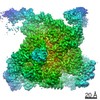
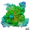
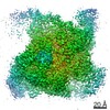
- PDB-7mkj: Cryo-EM structure of Escherichia coli RNA polymerase bound to T7A... -
+ Open data

- Basic information
| Entry | Database: PDB / ID: 7mkj | ||||||||||||||||||||||||||||||||||||||||||||||||||||||
|---|---|---|---|---|---|---|---|---|---|---|---|---|---|---|---|---|---|---|---|---|---|---|---|---|---|---|---|---|---|---|---|---|---|---|---|---|---|---|---|---|---|---|---|---|---|---|---|---|---|---|---|---|---|---|---|
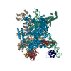
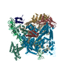
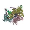
| Title | Cryo-EM structure of Escherichia coli RNA polymerase bound to T7A1 promoter DNA | ||||||||||||||||||||||||||||||||||||||||||||||||||||||
 Components Components |
| ||||||||||||||||||||||||||||||||||||||||||||||||||||||
 Keywords Keywords | TRANSCRIPTION/DNA / DNA-dependent RNA polymerase / transcription / DNA promoter / open complex / TRANSCRIPTION-DNA complex | ||||||||||||||||||||||||||||||||||||||||||||||||||||||
| Function / homology |  Function and homology information Function and homology informationsigma factor activity / DNA-directed RNA polymerase complex / DNA-templated transcription initiation / ribonucleoside binding / DNA-directed RNA polymerase / DNA-directed RNA polymerase activity / protein dimerization activity / DNA-templated transcription / magnesium ion binding / DNA binding ...sigma factor activity / DNA-directed RNA polymerase complex / DNA-templated transcription initiation / ribonucleoside binding / DNA-directed RNA polymerase / DNA-directed RNA polymerase activity / protein dimerization activity / DNA-templated transcription / magnesium ion binding / DNA binding / zinc ion binding / metal ion binding / cytoplasm Similarity search - Function | ||||||||||||||||||||||||||||||||||||||||||||||||||||||
| Biological species |    Escherichia phage T7 (virus) Escherichia phage T7 (virus) | ||||||||||||||||||||||||||||||||||||||||||||||||||||||
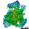
| Method | ELECTRON MICROSCOPY / single particle reconstruction / cryo EM / Resolution: 2.9 Å | ||||||||||||||||||||||||||||||||||||||||||||||||||||||
 Authors Authors | Saecker, R.M. / Darst, S.A. / Chen, J. | ||||||||||||||||||||||||||||||||||||||||||||||||||||||
| Funding support |  United States, 1items United States, 1items
| ||||||||||||||||||||||||||||||||||||||||||||||||||||||
 Citation Citation | Journal: Proc Natl Acad Sci U S A / Year: 2021 Title: Structural origins of RNA polymerase open promoter complex stability. Authors: Ruth M Saecker / James Chen / Courtney E Chiu / Brandon Malone / Johanna Sotiris / Mark Ebrahim / Laura Y Yen / Edward T Eng / Seth A Darst / Abstract: The first step in gene expression in all organisms requires opening the DNA duplex to expose one strand for templated RNA synthesis. In , promoter DNA sequence fundamentally determines how fast the ...The first step in gene expression in all organisms requires opening the DNA duplex to expose one strand for templated RNA synthesis. In , promoter DNA sequence fundamentally determines how fast the RNA polymerase (RNAP) forms "open" complexes (RPo), whether RPo persists for seconds or hours, and how quickly RNAP transitions from initiation to elongation. These rates control promoter strength in vivo, but their structural origins remain largely unknown. Here, we use cryoelectron microscopy to determine the structures of RPo formed de novo at three promoters with widely differing lifetimes at 37 °C: λP (t ∼10 h), T7A1 (t ∼4 min), and a point mutant in λP (λP) (t ∼2 h). Two distinct RPo conformers are populated at λP, likely representing productive and unproductive forms of RPo observed in solution studies. We find that changes in the sequence and length of DNA in the transcription bubble just upstream of the start site (+1) globally alter the network of DNA-RNAP interactions, base stacking, and strand order in the single-stranded DNA of the transcription bubble; these differences propagate beyond the bubble to upstream and downstream DNA. After expanding the transcription bubble by one base (T7A1), the nontemplate strand "scrunches" inside the active site cleft; the template strand bulges outside the cleft at the upstream edge of the bubble. The structures illustrate how limited sequence changes trigger global alterations in the transcription bubble that modulate the RPo lifetime and affect the subsequent steps of the transcription cycle. | ||||||||||||||||||||||||||||||||||||||||||||||||||||||
| History |
|
- Structure visualization
| Movie |
Movie viewer |
|---|---|
| Structure viewer | Molecule: MolmilJmol/JSmol |
- Downloads & links
-Download
| PDBx/mmCIF format | 7mkj.cif.gz | 730.1 KB | Display | PDBx/mmCIF format |
|---|---|---|---|---|
| PDB format | pdb7mkj.ent.gz | 577.8 KB | Display | PDB format |
| PDBx/mmJSON format | 7mkj.json.gz | Tree view | PDBx/mmJSON format | |
| Others |  Other downloads Other downloads |
-Validation report
| Arichive directory | https://data.pdbj.org/pub/pdb/validation_reports/mk/7mkjftp://data.pdbj.org/pub/pdb/validation_reports/mk/7mkj | HTTPS FTP |
|---|
-Related structure data
| Related structure data | 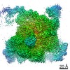 23897MC 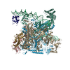 7mkdC 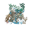 7mkeC 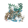 7mkiC M: map data used to model this data C: citing same article ( |
|---|---|
| Similar structure data |


-Links
 PDBj
PDBj


- Assembly
| Deposited unit | 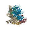
|
|---|---|
| 1 |
|
-Components
-DNA-directed RNA polymerase subunit ... , 4 types, 6 molecules GHRIJK
| #1: Protein | Mass: 36558.680 Da / Num. of mol.: 3 Source method: isolated from a genetically manipulated source Source: (gene. exp.) References: UniProt: A0A073G207, DNA-directed RNA polymerase #2: Protein | | Mass: 150820.875 Da / Num. of mol.: 1 Source method: isolated from a genetically manipulated source Source: (gene. exp.) #3: Protein | | Mass: 155366.781 Da / Num. of mol.: 1 Source method: isolated from a genetically manipulated source Source: (gene. exp.) References: UniProt: A0A4S1NBU2, DNA-directed RNA polymerase #4: Protein | | Mass: 10249.547 Da / Num. of mol.: 1 Source method: isolated from a genetically manipulated source Source: (gene. exp.) |
|---|
-Protein , 1 types, 1 molecules L
| #5: Protein | Mass: 70352.242 Da / Num. of mol.: 1 Source method: isolated from a genetically manipulated source Source: (gene. exp.) |
|---|
-DNA chain , 2 types, 2 molecules PQ
| #6: DNA chain | Mass: 28137.123 Da / Num. of mol.: 1 / Source method: obtained synthetically / Source: (synth.) Escherichia phage T7 (virus) / References: GenBank: 312369 |
|---|---|
| #7: DNA chain | Mass: 27988.918 Da / Num. of mol.: 1 / Source method: obtained synthetically / Source: (synth.) Escherichia phage T7 (virus) / References: GenBank: 312369 |
-Non-polymers , 3 types, 6 molecules 


| #8: Chemical |  Mass: 631.884 Da / Num. of mol.: 3 / Source method: obtained synthetically / Formula: C32H59N2O8S / Comment: detergent*YM Mass: 631.884 Da / Num. of mol.: 3 / Source method: obtained synthetically / Formula: C32H59N2O8S / Comment: detergent*YM#9: Chemical | ChemComp-MG / |  Mass: 24.305 Da / Num. of mol.: 1 / Source method: obtained synthetically / Formula: Mg Mass: 24.305 Da / Num. of mol.: 1 / Source method: obtained synthetically / Formula: Mg#10: Chemical |  Mass: 65.409 Da / Num. of mol.: 2 / Source method: obtained synthetically / Formula: Zn Mass: 65.409 Da / Num. of mol.: 2 / Source method: obtained synthetically / Formula: Zn |
|---|
-Details
| Has ligand of interest | N |
|---|---|
| Has protein modification | N |
-Experimental details
-Experiment
| Experiment | Method: ELECTRON MICROSCOPY |
|---|---|
| EM experiment | Aggregation state: PARTICLE / 3D reconstruction method: single particle reconstruction |
- Sample preparation
| Component |
| ||||||||||||||||||||||||
|---|---|---|---|---|---|---|---|---|---|---|---|---|---|---|---|---|---|---|---|---|---|---|---|---|---|
| Source (natural) |
| ||||||||||||||||||||||||
| Source (recombinant) | Organism: | ||||||||||||||||||||||||
| Buffer solution | pH: 8 | ||||||||||||||||||||||||
| Specimen | Embedding applied: NO / Shadowing applied: NO / Staining applied: NO / Vitrification applied: YES | ||||||||||||||||||||||||
| Vitrification | Cryogen name: ETHANE |
- Electron microscopy imaging
| Experimental equipment |  Model: Titan Krios / Image courtesy: FEI Company |
|---|---|
| Microscopy | Model: FEI TITAN KRIOS |
| Electron gun | Electron source:  FIELD EMISSION GUN / Accelerating voltage: 300 kV / Illumination mode: FLOOD BEAM FIELD EMISSION GUN / Accelerating voltage: 300 kV / Illumination mode: FLOOD BEAM |
| Electron lens | Mode: BRIGHT FIELD |
| Image recording | Electron dose: 43 e/Å2 / Film or detector model: GATAN K2 SUMMIT (4k x 4k) |
- Processing
| Software | Name: PHENIX / Version: 1.18.2_3874: / Classification: refinement | ||||||||||||||||||||||||
|---|---|---|---|---|---|---|---|---|---|---|---|---|---|---|---|---|---|---|---|---|---|---|---|---|---|
| EM software | Name: PHENIX / Category: model refinement | ||||||||||||||||||||||||
| CTF correction | Type: PHASE FLIPPING AND AMPLITUDE CORRECTION | ||||||||||||||||||||||||
| 3D reconstruction | Resolution: 2.9 Å / Resolution method: FSC 0.143 CUT-OFF / Num. of particles: 346991 / Symmetry type: POINT | ||||||||||||||||||||||||
| Refine LS restraints |
|
