Movie
Movie Controller


+ Open data

- Basic information
| Entry | Database: PDB / ID: 7miz | ||||||
|---|---|---|---|---|---|---|---|
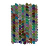
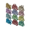
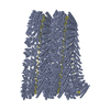
| Title | Atomic structure of cortical microtubule from Toxoplasma gondii | ||||||
 Components Components |
| ||||||
 Keywords Keywords | STRUCTURAL PROTEIN / cortical / parasite | ||||||
| Function / homology |  Function and homology information Function and homology informationthioredoxin-disulfide reductase (NADPH) activity / negative regulation of Wnt signaling pathway / microtubule-based process / negative regulation of protein ubiquitination / structural constituent of cytoskeleton / microtubule binding / microtubule / Hydrolases; Acting on acid anhydrides; Acting on GTP to facilitate cellular and subcellular movement / cytoskeleton / hydrolase activity ...thioredoxin-disulfide reductase (NADPH) activity / negative regulation of Wnt signaling pathway / microtubule-based process / negative regulation of protein ubiquitination / structural constituent of cytoskeleton / microtubule binding / microtubule / Hydrolases; Acting on acid anhydrides; Acting on GTP to facilitate cellular and subcellular movement / cytoskeleton / hydrolase activity / GTPase activity / GTP binding / metal ion binding / nucleus Similarity search - Function | ||||||
| Biological species |  | ||||||
| Method | ELECTRON MICROSCOPY / single particle reconstruction / cryo EM / Resolution: 3.4 Å | ||||||
 Authors Authors | Wang, X. / Brown, A. / Sibley, L.D. / Zhang, R. | ||||||
| Funding support |  United States, 1items United States, 1items
| ||||||
 Citation Citation | Journal: Nat Commun / Year: 2021 Title: Cryo-EM structure of cortical microtubules from human parasite Toxoplasma gondii identifies their microtubule inner proteins. Authors: Xiangli Wang / Yong Fu / Wandy L Beatty / Meisheng Ma / Alan Brown / L David Sibley / Rui Zhang / Abstract: In living cells, microtubules (MTs) play pleiotropic roles, which require very different mechanical properties. Unlike the dynamic MTs found in the cytoplasm of metazoan cells, the specialized ...In living cells, microtubules (MTs) play pleiotropic roles, which require very different mechanical properties. Unlike the dynamic MTs found in the cytoplasm of metazoan cells, the specialized cortical MTs from Toxoplasma gondii, a prevalent human pathogen, are extraordinarily stable and resistant to detergent and cold treatments. Using single-particle cryo-EM, we determine their ex vivo structure and identify three proteins (TrxL1, TrxL2 and SPM1) as bona fide microtubule inner proteins (MIPs). These three MIPs form a mesh on the luminal surface and simultaneously stabilize the tubulin lattice in both longitudinal and lateral directions. Consistent with previous observations, deletion of the identified MIPs compromises MT stability and integrity under challenges by chemical treatments. We also visualize a small molecule like density at the Taxol-binding site of β-tubulin. Our results provide the structural basis to understand the stability of cortical MTs and suggest an evolutionarily conserved mechanism of MT stabilization from the inside. | ||||||
| History |
|
- Structure visualization
| Movie |
Movie viewer |
|---|---|
| Structure viewer | Molecule: MolmilJmol/JSmol |
- Downloads & links
-Download
| PDBx/mmCIF format | 7miz.cif.gz | 5.1 MB | Display | PDBx/mmCIF format |
|---|---|---|---|---|
| PDB format | pdb7miz.ent.gz | Display | PDB format | |
| PDBx/mmJSON format | 7miz.json.gz | Tree view | PDBx/mmJSON format | |
| Others |  Other downloads Other downloads |
-Validation report
| Arichive directory | https://data.pdbj.org/pub/pdb/validation_reports/mi/7mizftp://data.pdbj.org/pub/pdb/validation_reports/mi/7miz | HTTPS FTP |
|---|
-Related structure data
| Related structure data |  23869MC M: map data used to model this data C: citing same article ( |
|---|---|
| Similar structure data |


-Links
 PDBj
PDBj


- Assembly
| Deposited unit | 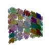
|
|---|---|
| 1 |
|
-Components
-Protein , 5 types, 100 molecules 01234567891011121314151617181920212223A0A2A4A6A8B0...
| #1: Protein | Mass: 38845.750 Da / Num. of mol.: 24 / Source method: isolated from a natural source / Source: (natural) #2: Protein | Mass: 50166.645 Da / Num. of mol.: 26 / Source method: isolated from a natural source / Source: (natural) #3: Protein | Mass: 50119.121 Da / Num. of mol.: 26 / Source method: isolated from a natural source / Source: (natural) #4: Protein | Mass: 24939.332 Da / Num. of mol.: 20 / Source method: isolated from a natural source / Source: (natural) #5: Protein | Mass: 21687.934 Da / Num. of mol.: 4 / Source method: isolated from a natural source / Source: (natural) |
|---|
-Non-polymers , 3 types, 78 molecules 


| #6: Chemical | ChemComp-GTP /  Mass: 523.180 Da / Num. of mol.: 26 / Source method: obtained synthetically / Formula: C10H16N5O14P3 / Comment: GTP, energy-carrying molecule*YM Mass: 523.180 Da / Num. of mol.: 26 / Source method: obtained synthetically / Formula: C10H16N5O14P3 / Comment: GTP, energy-carrying molecule*YM#7: Chemical | ChemComp-MG /  Mass: 24.305 Da / Num. of mol.: 26 / Source method: obtained synthetically / Formula: Mg Mass: 24.305 Da / Num. of mol.: 26 / Source method: obtained synthetically / Formula: Mg#8: Chemical | ChemComp-GDP /  Type: RNA linking / Mass: 443.201 Da / Num. of mol.: 26 / Source method: obtained synthetically / Formula: C10H15N5O11P2 / Comment: GDP, energy-carrying molecule*YM Type: RNA linking / Mass: 443.201 Da / Num. of mol.: 26 / Source method: obtained synthetically / Formula: C10H15N5O11P2 / Comment: GDP, energy-carrying molecule*YM |
|---|
-Details
| Has ligand of interest | N |
|---|---|
| Has protein modification | Y |
-Experimental details
-Experiment
| Experiment | Method: ELECTRON MICROSCOPY |
|---|---|
| EM experiment | Aggregation state: FILAMENT / 3D reconstruction method: single particle reconstruction |
- Sample preparation
| Component | Name: cortical microtubule with associated MIPs / Type: COMPLEX / Entity ID: #1-#5 / Source: NATURAL |
|---|---|
| Molecular weight | Experimental value: NO |
| Source (natural) | Organism: |
| Buffer solution | pH: 7.4 |
| Specimen | Embedding applied: NO / Shadowing applied: NO / Staining applied: NO / Vitrification applied: YES |
| Specimen support | Grid material: COPPER / Grid mesh size: 300 divisions/in. / Grid type: Quantifoil R2/2 |
| Vitrification | Instrument: FEI VITROBOT MARK IV / Cryogen name: ETHANE / Humidity: 95 % / Chamber temperature: 289 K / Details: blot for 4 seconds with a blot force of -15 |
- Electron microscopy imaging
| Experimental equipment |  Model: Titan Krios / Image courtesy: FEI Company |
|---|---|
| Microscopy | Model: FEI TITAN KRIOS |
| Electron gun | Electron source:  FIELD EMISSION GUN / Accelerating voltage: 300 kV / Illumination mode: FLOOD BEAM FIELD EMISSION GUN / Accelerating voltage: 300 kV / Illumination mode: FLOOD BEAM |
| Electron lens | Mode: BRIGHT FIELD / Nominal magnification: 105000 X / Nominal defocus max: 3500 nm / Calibrated defocus min: 1000 nm / Cs: 0.01 mm / Alignment procedure: ZEMLIN TABLEAU |
| Specimen holder | Cryogen: NITROGEN / Specimen holder model: FEI TITAN KRIOS AUTOGRID HOLDER |
| Image recording | Average exposure time: 9 sec. / Electron dose: 63.7 e/Å2 / Film or detector model: GATAN K3 BIOQUANTUM (6k x 4k) / Num. of grids imaged: 2 / Num. of real images: 9231 Details: Images were collected in counting mode, with an exposure rate of 8.5 electrons/pixel/s on the detector camera. |
| Image scans | Width: 3838 / Height: 3710 |
- Processing
| EM software |
| ||||||||||||||||||||||||||||||||||||||||||||||||||||
|---|---|---|---|---|---|---|---|---|---|---|---|---|---|---|---|---|---|---|---|---|---|---|---|---|---|---|---|---|---|---|---|---|---|---|---|---|---|---|---|---|---|---|---|---|---|---|---|---|---|---|---|---|---|
| CTF correction | Details: CTF amplitude correction was performed as part of the 3D reconstruction. Type: PHASE FLIPPING AND AMPLITUDE CORRECTION | ||||||||||||||||||||||||||||||||||||||||||||||||||||
| Particle selection | Num. of particles selected: 536132 Details: Cortical MTs were manually picked from the motion-corrected micrographs using the APPION image-processing suite. The selected MT segments were computationally cut into overlapping boxes ...Details: Cortical MTs were manually picked from the motion-corrected micrographs using the APPION image-processing suite. The selected MT segments were computationally cut into overlapping boxes (512x512 pixels) with an 8-nm non-overlapping region (step size) between adjacent boxes (corresponding to the length of a tubulin heterodimer). We refer to these boxed images as 8-nm MT particles. | ||||||||||||||||||||||||||||||||||||||||||||||||||||
| Symmetry | Point symmetry: C1 (asymmetric) | ||||||||||||||||||||||||||||||||||||||||||||||||||||
| 3D reconstruction | Resolution: 3.4 Å / Resolution method: FSC 0.143 CUT-OFF / Num. of particles: 220139 / Algorithm: FOURIER SPACE / Symmetry type: POINT | ||||||||||||||||||||||||||||||||||||||||||||||||||||
| Atomic model building | Protocol: AB INITIO MODEL Details: Secondary structure, Ramachandran, and rotamer restraints were applied during refinement. The nonbonded_weight value was set to 500 to reduce the clashes between atoms. | ||||||||||||||||||||||||||||||||||||||||||||||||||||
| Atomic model building |
|