 Movie
Movie Controller


+ Open data

- Basic information
| Entry | Database: PDB / ID: 7mff | |||||||||
|---|---|---|---|---|---|---|---|---|---|---|
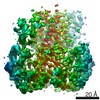
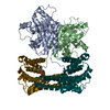
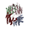
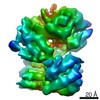
| Title | Dimeric (BRAF)2:(14-3-3)2 complex bound to SB590885 Inhibitor | |||||||||
 Components Components |
| |||||||||
 Keywords Keywords | SIGNALING PROTEIN / B-Raf / 14-3-3 / B-Raf complex / B-Raf dimer / Active B-Raf / SB590885 / Serine/threonine-protein kinase B-raf | |||||||||
| Function / homology |  Function and homology information Function and homology informationsynaptic target recognition / Golgi reassembly / NOTCH4 Activation and Transmission of Signal to the Nucleus / respiratory system process / establishment of Golgi localization / tube formation / Signalling to p38 via RIT and RIN / ARMS-mediated activation / regulation of synapse maturation / SHOC2 M1731 mutant abolishes MRAS complex function ...synaptic target recognition / Golgi reassembly / NOTCH4 Activation and Transmission of Signal to the Nucleus / respiratory system process / establishment of Golgi localization / tube formation / Signalling to p38 via RIT and RIN / ARMS-mediated activation / regulation of synapse maturation / SHOC2 M1731 mutant abolishes MRAS complex function / Gain-of-function MRAS complexes activate RAF signaling / Rap1 signalling / positive regulation of D-glucose transmembrane transport / establishment of protein localization to membrane / negative regulation of protein localization to nucleus / KSRP (KHSRP) binds and destabilizes mRNA / Negative feedback regulation of MAPK pathway / GP1b-IX-V activation signalling / Frs2-mediated activation / MAP kinase kinase activity / lung development / Regulation of localization of FOXO transcription factors / Interleukin-3, Interleukin-5 and GM-CSF signaling / positive regulation of peptidyl-serine phosphorylation / phosphoserine residue binding / Activation of BAD and translocation to mitochondria / regulation of ERK1 and ERK2 cascade / MAP kinase kinase kinase activity / postsynaptic modulation of chemical synaptic transmission / cellular response to glucose starvation / SARS-CoV-2 targets host intracellular signalling and regulatory pathways / SARS-CoV-1 targets host intracellular signalling and regulatory pathways / RHO GTPases activate PKNs / Chk1/Chk2(Cds1) mediated inactivation of Cyclin B:Cdk1 complex / negative regulation of TORC1 signaling / animal organ morphogenesis / Transcriptional and post-translational regulation of MITF-M expression and activity / cellular response to calcium ion / negative regulation of innate immune response / hippocampal mossy fiber to CA3 synapse / TP53 Regulates Metabolic Genes / Translocation of SLC2A4 (GLUT4) to the plasma membrane / regulation of protein stability / RAF activation / protein sequestering activity / Signaling by high-kinase activity BRAF mutants / Spry regulation of FGF signaling / Deactivation of the beta-catenin transactivating complex / MAP2K and MAPK activation / Negative regulation of NOTCH4 signaling / epidermal growth factor receptor signaling pathway / intracellular protein localization / Signaling by RAF1 mutants / Signaling by moderate kinase activity BRAF mutants / Paradoxical activation of RAF signaling by kinase inactive BRAF / Signaling downstream of RAS mutants / MAPK cascade / Negative regulation of MAPK pathway / Signaling by BRAF and RAF1 fusions / melanosome / angiogenesis / cell body / scaffold protein binding / protein phosphatase binding / blood microparticle / DNA-binding transcription factor binding / vesicle / positive regulation of ERK1 and ERK2 cascade / transmembrane transporter binding / protein kinase activity / protein phosphorylation / non-specific serine/threonine protein kinase / postsynapse / neuron projection / cadherin binding / protein domain specific binding / protein serine kinase activity / focal adhesion / ubiquitin protein ligase binding / protein serine/threonine kinase activity / positive regulation of gene expression / calcium ion binding / negative regulation of apoptotic process / protein kinase binding / negative regulation of transcription by RNA polymerase II / glutamatergic synapse / signal transduction / mitochondrion / : / RNA binding / extracellular exosome / nucleoplasm / zinc ion binding / ATP binding / identical protein binding / nucleus / plasma membrane / cytosol / cytoplasm Similarity search - Function | |||||||||
| Biological species |  Homo sapiens (human) Homo sapiens (human) | |||||||||
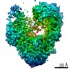
| Method | ELECTRON MICROSCOPY / single particle reconstruction / cryo EM / Resolution: 3.89 Å | |||||||||
 Authors Authors | Martinez Fiesco, J.A. / Ping, Z. / Durrant, D.E. / Morrison, D.K. | |||||||||
| Funding support |  United States, 2items United States, 2items
| |||||||||
 Citation Citation | Journal: Nat Commun / Year: 2022 Title: Structural insights into the BRAF monomer-to-dimer transition mediated by RAS binding. Authors: Juliana A Martinez Fiesco / David E Durrant / Deborah K Morrison / Ping Zhang / Abstract: RAF kinases are essential effectors of RAS, but how RAS binding initiates the conformational changes needed for autoinhibited RAF monomers to form active dimers has remained unclear. Here, we present ...RAF kinases are essential effectors of RAS, but how RAS binding initiates the conformational changes needed for autoinhibited RAF monomers to form active dimers has remained unclear. Here, we present cryo-electron microscopy structures of full-length BRAF complexes derived from mammalian cells: autoinhibited, monomeric BRAF:14-3-3:MEK and BRAF:14-3-3 complexes, and an inhibitor-bound, dimeric BRAF:14-3-3 complex, at 3.7, 4.1, and 3.9 Å resolution, respectively. In both autoinhibited, monomeric structures, the RAS binding domain (RBD) of BRAF is resolved, revealing that the RBD forms an extensive contact interface with the 14-3-3 protomer bound to the BRAF C-terminal site and that key basic residues required for RBD-RAS binding are exposed. Moreover, through structure-guided mutational studies, our findings indicate that RAS-RAF binding is a dynamic process and that RBD residues at the center of the RBD:14-3-3 interface have a dual function, first contributing to RAF autoinhibition and then to the full spectrum of RAS-RBD interactions. | |||||||||
| History |
|
- Structure visualization
| Movie |
Movie viewer |
|---|---|
| Structure viewer | Molecule: MolmilJmol/JSmol |
- Downloads & links
-Download
| PDBx/mmCIF format | 7mff.cif.gz | 201.8 KB | Display | PDBx/mmCIF format |
|---|---|---|---|---|
| PDB format | pdb7mff.ent.gz | 151.4 KB | Display | PDB format |
| PDBx/mmJSON format | 7mff.json.gz | Tree view | PDBx/mmJSON format | |
| Others |  Other downloads Other downloads |
-Validation report
| Arichive directory | https://data.pdbj.org/pub/pdb/validation_reports/mf/7mffftp://data.pdbj.org/pub/pdb/validation_reports/mf/7mff | HTTPS FTP |
|---|
-Related structure data
| Related structure data | 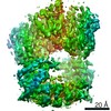 23815MC 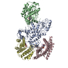 7mfdC 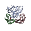 7mfeC M: map data used to model this data C: citing same article ( |
|---|---|
| Similar structure data |


-Links
 PDBj
PDBj



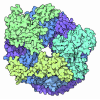




- Assembly
| Deposited unit | 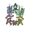
|
|---|---|
| 1 |
|
-Components
| #1: Protein | Mass: 27777.092 Da / Num. of mol.: 2 Source method: isolated from a genetically manipulated source Source: (gene. exp.) Homo sapiens (human) / Production host: Homo sapiens (human) / References: UniProt: P63104#2: Protein | Mass: 84697.695 Da / Num. of mol.: 2 Source method: isolated from a genetically manipulated source Source: (gene. exp.) Homo sapiens (human) / Gene: BRAF, BRAF1, RAFB1 / Production host: Homo sapiens (human)References: UniProt: P15056, non-specific serine/threonine protein kinase #3: Chemical |  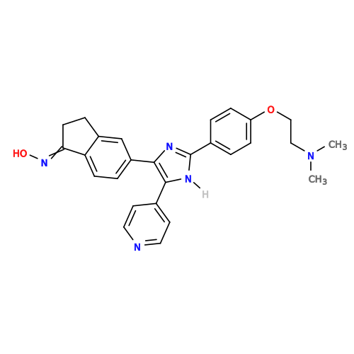 Mass: 453.536 Da / Num. of mol.: 2 / Source method: obtained synthetically / Formula: C27H27N5O2 Mass: 453.536 Da / Num. of mol.: 2 / Source method: obtained synthetically / Formula: C27H27N5O2Has ligand of interest | N | Has protein modification | Y | |
|---|
-Experimental details
-Experiment
| Experiment | Method: ELECTRON MICROSCOPY |
|---|---|
| EM experiment | Aggregation state: PARTICLE / 3D reconstruction method: single particle reconstruction |
- Sample preparation
| Component | Name: Active dimeric (B-Raf)2:(14-3-3)2 complex / Type: COMPLEX / Entity ID: #1-#2 / Source: RECOMBINANT | ||||||||||||||||
|---|---|---|---|---|---|---|---|---|---|---|---|---|---|---|---|---|---|
| Molecular weight | Experimental value: NO | ||||||||||||||||
| Source (natural) | Organism: Homo sapiens (human) | ||||||||||||||||
| Buffer solution | pH: 8 | ||||||||||||||||
| Buffer component |
| ||||||||||||||||
| Specimen | Conc.: 0.2 mg/ml / Embedding applied: NO / Shadowing applied: NO / Staining applied: NO / Vitrification applied: YES | ||||||||||||||||
| Vitrification | Instrument: FEI VITROBOT MARK IV / Cryogen name: ETHANE / Humidity: 90 % / Chamber temperature: 277.15 K |
- Electron microscopy imaging
| Experimental equipment |  Model: Titan Krios / Image courtesy: FEI Company |
|---|---|
| Microscopy | Model: FEI TITAN KRIOS |
| Electron gun | Electron source:  FIELD EMISSION GUN / Accelerating voltage: 300 kV / Illumination mode: FLOOD BEAM FIELD EMISSION GUN / Accelerating voltage: 300 kV / Illumination mode: FLOOD BEAM |
| Electron lens | Mode: BRIGHT FIELD |
| Image recording | Electron dose: 57 e/Å2 / Detector mode: SUPER-RESOLUTION / Film or detector model: GATAN K2 SUMMIT (4k x 4k) |
- Processing
| Software | Name: PHENIX / Version: 1.19.2_4158: / Classification: refinement | ||||||||||||||||||||||||
|---|---|---|---|---|---|---|---|---|---|---|---|---|---|---|---|---|---|---|---|---|---|---|---|---|---|
| CTF correction | Type: PHASE FLIPPING AND AMPLITUDE CORRECTION | ||||||||||||||||||||||||
| 3D reconstruction | Resolution: 3.89 Å / Resolution method: FSC 0.143 CUT-OFF / Num. of particles: 203343 / Symmetry type: POINT | ||||||||||||||||||||||||
| Refine LS restraints |
|
