 ムービー
ムービー コントローラー


+ データを開く

- 基本情報
基本情報
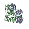
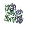
| 登録情報 | データベース: PDB / ID: 7m4r | ||||||||||||||||||||||||||||||||||||
|---|---|---|---|---|---|---|---|---|---|---|---|---|---|---|---|---|---|---|---|---|---|---|---|---|---|---|---|---|---|---|---|---|---|---|---|---|---|
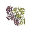
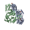
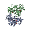
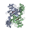
| タイトル | Structural basis for SARS-CoV-2 envelope protein in recognition of human cell junction protein PALS1 | ||||||||||||||||||||||||||||||||||||
 要素 要素 |
| ||||||||||||||||||||||||||||||||||||
 キーワード キーワード | CELL ADHESION/VIRAL PROTEIN / SARS-CoV-2 envelope protein / PDZ-binding motif / complex / pathogen-host interaction / CELL ADHESION-VIRAL PROTEIN complex | ||||||||||||||||||||||||||||||||||||
| 機能・相同性 |  機能・相同性情報 機能・相同性情報protein localization to myelin sheath abaxonal region / disruption of cellular anatomical structure in another organism / SARS-CoV-1 targets PDZ proteins in cell-cell junction / symbiont-mediated perturbation of host cell endomembrane system / viral budding from Golgi membrane / myelin assembly / establishment or maintenance of polarity of embryonic epithelium / morphogenesis of an epithelial sheet / Tight junction interactions / SARS-CoV-2 targets PDZ proteins in cell-cell junction ...protein localization to myelin sheath abaxonal region / disruption of cellular anatomical structure in another organism / SARS-CoV-1 targets PDZ proteins in cell-cell junction / symbiont-mediated perturbation of host cell endomembrane system / viral budding from Golgi membrane / myelin assembly / establishment or maintenance of polarity of embryonic epithelium / morphogenesis of an epithelial sheet / Tight junction interactions / SARS-CoV-2 targets PDZ proteins in cell-cell junction / regulation of transforming growth factor beta receptor signaling pathway / myelin sheath adaxonal region / Regulation of gap junction activity / lateral loop / Schmidt-Lanterman incisure / establishment or maintenance of epithelial cell apical/basal polarity / peripheral nervous system myelin maintenance / apical junction complex / generation of neurons / central nervous system neuron development / host cell Golgi membrane / bicellular tight junction / endoplasmic reticulum-Golgi intermediate compartment membrane / Maturation of protein E / protein localization to plasma membrane / adherens junction / cerebral cortex development / Translation of Structural Proteins / Virion Assembly and Release / Induction of Cell-Cell Fusion / gene expression / perikaryon / Attachment and Entry / apical plasma membrane / protein domain specific binding / axon / SARS-CoV-2 activates/modulates innate and adaptive immune responses / virion membrane / Golgi apparatus / protein-containing complex / extracellular exosome / ATP binding / membrane / identical protein binding / plasma membrane / cytoplasm 類似検索 - 分子機能 | ||||||||||||||||||||||||||||||||||||
| 生物種 |  Homo sapiens (ヒト) Homo sapiens (ヒト)  Severe acute respiratory syndrome coronavirus 2 (ウイルス) Severe acute respiratory syndrome coronavirus 2 (ウイルス) | ||||||||||||||||||||||||||||||||||||
| 手法 | 電子顕微鏡法 / 単粒子再構成法 / クライオ電子顕微鏡法 / 解像度: 3.65 Å | ||||||||||||||||||||||||||||||||||||
 データ登録者 データ登録者 | Liu, Q. / Chai, J. | ||||||||||||||||||||||||||||||||||||
 引用 引用 | ジャーナル: Nat Commun / 年: 2021 タイトル: Structural basis for SARS-CoV-2 envelope protein recognition of human cell junction protein PALS1. 著者: Jin Chai / Yuanheng Cai / Changxu Pang / Liguo Wang / Sean McSweeney / John Shanklin / Qun Liu /  要旨: The COVID-19 pandemic, caused by the SARS-CoV-2 virus, has created global health and economic emergencies. SARS-CoV-2 viruses promote their own spread and virulence by hijacking human proteins, which ...The COVID-19 pandemic, caused by the SARS-CoV-2 virus, has created global health and economic emergencies. SARS-CoV-2 viruses promote their own spread and virulence by hijacking human proteins, which occurs through viral protein recognition of human targets. To understand the structural basis for SARS-CoV-2 viral-host protein recognition, here we use cryo-electron microscopy (cryo-EM) to determine a complex structure of the human cell junction protein PALS1 and SARS-CoV-2 viral envelope (E) protein. Our reported structure shows that the E protein C-terminal DLLV motif recognizes a pocket formed exclusively by hydrophobic residues from the PDZ and SH3 domains of PALS1. Our structural analysis provides an explanation for the observation that the viral E protein recruits PALS1 from lung epithelial cell junctions. In addition, our structure provides novel targets for peptide- and small-molecule inhibitors that could block the PALS1-E interactions to reduce E-mediated virulence. | ||||||||||||||||||||||||||||||||||||
| 履歴 |
|
- 構造の表示
構造の表示
| ムービー |
ムービービューア |
|---|---|
| 構造ビューア | 分子: MolmilJmol/JSmol |
- ダウンロードとリンク
-ダウンロード
| PDBx/mmCIF形式 | 7m4r.cif.gz | 165 KB | 表示 | PDBx/mmCIF形式 |
|---|---|---|---|---|
| PDB形式 | pdb7m4r.ent.gz | 120.7 KB | 表示 | PDB形式 |
| PDBx/mmJSON形式 | 7m4r.json.gz | ツリー表示 | PDBx/mmJSON形式 | |
| その他 |  その他のダウンロード その他のダウンロード |
-検証レポート
| アーカイブディレクトリ | https://data.pdbj.org/pub/pdb/validation_reports/m4/7m4rftp://data.pdbj.org/pub/pdb/validation_reports/m4/7m4r | HTTPS FTP |
|---|
-関連構造データ


-リンク
 PDBj
PDBj


- 集合体
集合体
| 登録構造単位 | 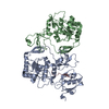
|
|---|---|
| 1 |
|
-要素
| #1: タンパク質 | 分子量: 44780.504 Da / 分子数: 2 / 断片: UNP residues 236-410,461-675 / 由来タイプ: 組換発現 / 由来: (組換発現) Homo sapiens (ヒト) / 遺伝子: MPP5, PALS1 / 発現宿主:  #2: タンパク質・ペプチド | | 分子量: 2062.395 Da / 分子数: 1 / 断片: UNP residues 58-75 / 由来タイプ: 合成 由来: (合成) Severe acute respiratory syndrome coronavirus 2 (ウイルス)参照: UniProt: P0DTC4 Has protein modification | N | |
|---|
-実験情報
-実験
| 実験 | 手法: 電子顕微鏡法 |
|---|---|
| EM実験 | 試料の集合状態: PARTICLE / 3次元再構成法: 単粒子再構成法 |
- 試料調製
試料調製
| 構成要素 | 名称: Complex structure of SARS-CoV-2 envelope protein Ec18 and human PALS1 PSG domains タイプ: COMPLEX / Entity ID: all / 由来: MULTIPLE SOURCES |
|---|---|
| 由来(天然) | 生物種: Homo sapiens (ヒト) |
| 由来(組換発現) | 生物種: |
| 緩衝液 | pH: 7.5 |
| 試料 | 包埋: NO / シャドウイング: NO / 染色: NO / 凍結: YES |
| 急速凍結 | 凍結剤: ETHANE |
- 電子顕微鏡撮影
| 実験機器 |  モデル: Titan Krios / 画像提供: FEI Company |
|---|---|
| 顕微鏡 | モデル: FEI TITAN KRIOS |
| 電子銃 | 電子線源:  FIELD EMISSION GUN / 加速電圧: 300 kV / 照射モード: FLOOD BEAM FIELD EMISSION GUN / 加速電圧: 300 kV / 照射モード: FLOOD BEAM |
| 電子レンズ | モード: BRIGHT FIELD |
| 撮影 | 電子線照射量: 64 e/Å2 フィルム・検出器のモデル: GATAN K3 BIOQUANTUM (6k x 4k) |
- 解析
解析
| ソフトウェア | 名称: PHENIX / バージョン: 1.18.2_3874: / 分類: 精密化 | ||||||||||||||||||||||||
|---|---|---|---|---|---|---|---|---|---|---|---|---|---|---|---|---|---|---|---|---|---|---|---|---|---|
| EMソフトウェア | 名称: PHENIX / カテゴリ: モデル精密化 | ||||||||||||||||||||||||
| CTF補正 | タイプ: PHASE FLIPPING AND AMPLITUDE CORRECTION | ||||||||||||||||||||||||
| 3次元再構成 | 解像度: 3.65 Å / 解像度の算出法: FSC 0.143 CUT-OFF / 粒子像の数: 47615 / 対称性のタイプ: POINT | ||||||||||||||||||||||||
| 拘束条件 |
|
