 ムービー
ムービー コントローラー


+ データを開く

- 基本情報
基本情報
| 登録情報 | データベース: PDB / ID: 7l9p | ||||||
|---|---|---|---|---|---|---|---|
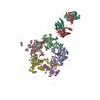
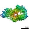
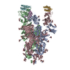
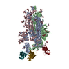
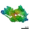
| タイトル | Structure of human SHLD2-SHLD3-REV7-TRIP13(E253Q) complex | ||||||
 要素 要素 |
| ||||||
 キーワード キーワード | NUCLEAR PROTEIN / REV7 / SHLD2 / SHLD3 / TRIP13 | ||||||
| 機能・相同性 |  機能・相同性情報 機能・相同性情報somatic diversification of immunoglobulins involved in immune response / DNA damage response, signal transduction resulting in transcription / negative regulation of transcription regulatory region DNA binding / meiotic recombination checkpoint signaling / zeta DNA polymerase complex / synaptonemal complex assembly / telomere maintenance in response to DNA damage / positive regulation of isotype switching / positive regulation of extracellular matrix assembly / negative regulation of transcription by competitive promoter binding ...somatic diversification of immunoglobulins involved in immune response / DNA damage response, signal transduction resulting in transcription / negative regulation of transcription regulatory region DNA binding / meiotic recombination checkpoint signaling / zeta DNA polymerase complex / synaptonemal complex assembly / telomere maintenance in response to DNA damage / positive regulation of isotype switching / positive regulation of extracellular matrix assembly / negative regulation of transcription by competitive promoter binding / negative regulation of cell-cell adhesion mediated by cadherin / JUN kinase binding / negative regulation of epithelial to mesenchymal transition / reciprocal meiotic recombination / female meiosis I / oocyte maturation / negative regulation of ubiquitin protein ligase activity / regulation of double-strand break repair via homologous recombination / oogenesis / mitotic spindle assembly checkpoint signaling / positive regulation of double-strand break repair via nonhomologous end joining / male meiosis I / spermatid development / negative regulation of double-strand break repair via homologous recombination / male germ cell nucleus / error-prone translesion synthesis / positive regulation of epithelial to mesenchymal transition / transcription coregulator activity / Translesion synthesis by REV1 / Translesion synthesis by POLK / Translesion synthesis by POLI / actin filament organization / negative regulation of protein catabolic process / regulation of cell growth / negative regulation of canonical Wnt signaling pathway / negative regulation of DNA-binding transcription factor activity / spindle / chromosome / positive regulation of peptidyl-serine phosphorylation / double-strand break repair / actin cytoskeleton / site of double-strand break / spermatogenesis / RNA polymerase II-specific DNA-binding transcription factor binding / transcription by RNA polymerase II / cell division / DNA repair / positive regulation of DNA-templated transcription / chromatin / nucleolus / negative regulation of transcription by RNA polymerase II / ATP hydrolysis activity / nucleoplasm / ATP binding / identical protein binding / nucleus / cytoplasm 類似検索 - 分子機能 | ||||||
| 生物種 |  Homo sapiens (ヒト) Homo sapiens (ヒト) | ||||||
| 手法 | 電子顕微鏡法 / 単粒子再構成法 / クライオ電子顕微鏡法 / 解像度: 3.6 Å | ||||||
 データ登録者 データ登録者 | Xie, W. / Patel, D.J. | ||||||
 引用 引用 | ジャーナル: Proc Natl Acad Sci U S A / 年: 2021 タイトル: Molecular mechanisms of assembly and TRIP13-mediated remodeling of the human Shieldin complex. 著者: Wei Xie / Shengliu Wang / Juncheng Wang / M Jason de la Cruz / Guotai Xu / Maurizio Scaltriti / Dinshaw J Patel /  要旨: The Shieldin complex, composed of REV7, SHLD1, SHLD2, and SHLD3, protects DNA double-strand breaks (DSBs) to promote nonhomologous end joining. The AAA ATPase TRIP13 remodels Shieldin to regulate DNA ...The Shieldin complex, composed of REV7, SHLD1, SHLD2, and SHLD3, protects DNA double-strand breaks (DSBs) to promote nonhomologous end joining. The AAA ATPase TRIP13 remodels Shieldin to regulate DNA repair pathway choice. Here we report crystal structures of human SHLD3-REV7 binary and fused SHLD2-SHLD3-REV7 ternary complexes, revealing that assembly of Shieldin requires fused SHLD2-SHLD3 induced conformational heterodimerization of open (O-REV7) and closed (C-REV7) forms of REV7. We also report the cryogenic electron microscopy (cryo-EM) structures of the ATPγS-bound fused SHLD2-SHLD3-REV7-TRIP13 complexes, uncovering the principles underlying the TRIP13-mediated disassembly mechanism of the Shieldin complex. We demonstrate that the N terminus of REV7 inserts into the central channel of TRIP13, setting the stage for pulling the unfolded N-terminal peptide of C-REV7 through the central TRIP13 hexameric channel. The primary interface involves contacts between the safety-belt segment of C-REV7 and a conserved and negatively charged loop of TRIP13. This process is mediated by ATP hydrolysis-triggered rotatory motions of the TRIP13 ATPase, thereby resulting in the disassembly of the Shieldin complex. | ||||||
| 履歴 |
|
- 構造の表示
構造の表示
| ムービー |
ムービービューア |
|---|---|
| 構造ビューア | 分子: MolmilJmol/JSmol |
- ダウンロードとリンク
-ダウンロード
| PDBx/mmCIF形式 | 7l9p.cif.gz | 545.2 KB | 表示 | PDBx/mmCIF形式 |
|---|---|---|---|---|
| PDB形式 | pdb7l9p.ent.gz | 435.6 KB | 表示 | PDB形式 |
| PDBx/mmJSON形式 | 7l9p.json.gz | ツリー表示 | PDBx/mmJSON形式 | |
| その他 |  その他のダウンロード その他のダウンロード |
-検証レポート
| 文書・要旨 | 7l9p_validation.pdf.gz | 1.3 MB | 表示 | wwPDB検証レポート |
|---|---|---|---|---|
| 文書・詳細版 | 7l9p_full_validation.pdf.gz | 1.4 MB | 表示 | |
| XML形式データ | 7l9p_validation.xml.gz | 88.1 KB | 表示 | |
| CIF形式データ | 7l9p_validation.cif.gz | 130.8 KB | 表示 | |
| アーカイブディレクトリ | https://data.pdbj.org/pub/pdb/validation_reports/l9/7l9pftp://data.pdbj.org/pub/pdb/validation_reports/l9/7l9p | HTTPS FTP |
-関連構造データ


-リンク
 PDBj
PDBj


- 集合体
集合体
| 登録構造単位 | 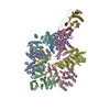
|
|---|---|
| 1 |
|
-要素
| #1: タンパク質 | 分子量: 48562.547 Da / 分子数: 6 / 変異: E253Q / 由来タイプ: 組換発現 / 由来: (組換発現) Homo sapiens (ヒト) / 遺伝子: TRIP13, PCH2 / 発現宿主:  #2: タンパク質 | 分子量: 24323.348 Da / 分子数: 4 / 由来タイプ: 組換発現 / 詳細: closed form / 由来: (組換発現) Homo sapiens (ヒト) / 遺伝子: MAD2L2, MAD2B, REV7 / 発現宿主: #3: タンパク質 | 分子量: 10678.139 Da / 分子数: 2 Fragment: SHLD2 (UNP residues 5-19) + linker + SHLD3 (UNP residues 2-58) 由来タイプ: 組換発現 / 由来: (組換発現) Homo sapiens (ヒト) / 遺伝子: SHLD2, FAM35A, RINN2, SHLD3, FLJ26957, RINN1 / 発現宿主: #4: 化合物 | ChemComp-AGS /   分子量: 523.247 Da / 分子数: 5 / 由来タイプ: 合成 / 式: C10H16N5O12P3S / タイプ: SUBJECT OF INVESTIGATION 分子量: 523.247 Da / 分子数: 5 / 由来タイプ: 合成 / 式: C10H16N5O12P3S / タイプ: SUBJECT OF INVESTIGATIONコメント: ATP-gamma-S, エネルギー貯蔵分子類似体*YM 研究の焦点であるリガンドがあるか | Y | |
|---|
-実験情報
-実験
| 実験 | 手法: 電子顕微鏡法 |
|---|---|
| EM実験 | 試料の集合状態: PARTICLE / 3次元再構成法: 単粒子再構成法 |
- 試料調製
試料調製
| 構成要素 | 名称: SHLD2.3-REV7(4)-TRIP13(E253Q) complex with ATP-gamma-S タイプ: COMPLEX / Entity ID: #1-#3 / 由来: RECOMBINANT |
|---|---|
| 由来(天然) | 生物種: Homo sapiens (ヒト) |
| 由来(組換発現) | 生物種: |
| 緩衝液 | pH: 7.3 詳細: 20 mM HEPES, pH 7.3, 300 mM NaCl, 5 mM MgCl2, 0.1 mM ATP-gamma-S, 1 mM DTT |
| 試料 | 濃度: 0.3 mg/ml / 包埋: NO / シャドウイング: NO / 染色: NO / 凍結: YES |
| 試料支持 | グリッドの材料: GOLD / グリッドのタイプ: UltrAuFoil R1.2/1.3 |
| 急速凍結 | 装置: FEI VITROBOT MARK IV / 凍結剤: ETHANE / 湿度: 100 % / 凍結前の試料温度: 277 K / 詳細: 1.5-second blot, blot force of 0 |
- 電子顕微鏡撮影
| 実験機器 |  モデル: Titan Krios / 画像提供: FEI Company |
|---|---|
| 顕微鏡 | モデル: FEI TITAN KRIOS |
| 電子銃 | 電子線源:  FIELD EMISSION GUN / 加速電圧: 300 kV / 照射モード: FLOOD BEAM FIELD EMISSION GUN / 加速電圧: 300 kV / 照射モード: FLOOD BEAM |
| 電子レンズ | モード: BRIGHT FIELD / 最大 デフォーカス(公称値): -2500 nm / 最小 デフォーカス(公称値): -1000 nm |
| 試料ホルダ | 凍結剤: NITROGEN 試料ホルダーモデル: FEI TITAN KRIOS AUTOGRID HOLDER |
| 撮影 | 平均露光時間: 0.075 sec. / 電子線照射量: 53 e/Å2 フィルム・検出器のモデル: GATAN K3 BIOQUANTUM (6k x 4k) 撮影したグリッド数: 1 / 実像数: 40 |
- 解析
解析
| ソフトウェア | 名称: PHENIX / バージョン: 1.18_3855: / 分類: 精密化 | ||||||||||||||||||||||||
|---|---|---|---|---|---|---|---|---|---|---|---|---|---|---|---|---|---|---|---|---|---|---|---|---|---|
| CTF補正 | タイプ: PHASE FLIPPING AND AMPLITUDE CORRECTION | ||||||||||||||||||||||||
| 対称性 | 点対称性: C2 (2回回転対称) | ||||||||||||||||||||||||
| 3次元再構成 | 解像度: 3.6 Å / 解像度の算出法: FSC 0.143 CUT-OFF / 粒子像の数: 104023 / 対称性のタイプ: POINT | ||||||||||||||||||||||||
| 拘束条件 |
|
