 Movie
Movie Controller

[English] 日本語
 Yorodumi
- PDB-7kdv: Murine core lysosomal multienzyme complex (LMC) composed of acid ... -
+ Open data

- Basic information
| Entry | Database: PDB / ID: 7kdv | ||||||||||||
|---|---|---|---|---|---|---|---|---|---|---|---|---|---|
| Title | Murine core lysosomal multienzyme complex (LMC) composed of acid beta-galactosidase (GLB1) and protective protein cathepsin A (PPCA, CTSA) | ||||||||||||
 Components Components |
| ||||||||||||
 Keywords Keywords | HYDROLASE / glycosidase / protease / lysosome | ||||||||||||
| Function / homology |  Function and homology information Function and homology informationKeratan sulfate degradation / HS-GAG degradation / keratan sulfate proteoglycan catabolic process / response to cortisone / Sialic acid metabolism / response to Thyroglobulin triiodothyronine / carboxypeptidase C / Glycosphingolipid catabolism / galactose catabolic process / serine-type carboxypeptidase activity ...Keratan sulfate degradation / HS-GAG degradation / keratan sulfate proteoglycan catabolic process / response to cortisone / Sialic acid metabolism / response to Thyroglobulin triiodothyronine / carboxypeptidase C / Glycosphingolipid catabolism / galactose catabolic process / serine-type carboxypeptidase activity / galactoside binding / glycoprotein catabolic process / beta-galactosidase / MHC class II antigen presentation / ganglioside catabolic process / beta-galactosidase activity / negative regulation of chaperone-mediated autophagy / Neutrophil degranulation / regulation of protein stability / lysosome / hydrolase activity / Golgi apparatus / protein homodimerization activity / mitochondrion / proteolysis / extracellular space / membrane / cytoplasm Similarity search - Function | ||||||||||||
| Biological species |  | ||||||||||||
| Method | ELECTRON MICROSCOPY / single particle reconstruction / cryo EM / Resolution: 4.59 Å | ||||||||||||
 Authors Authors | Gorelik, A. / Illes, K. / Hasan, S.M.N. / Nagar, B. / Mazhab-Jafari, M.T. | ||||||||||||
| Funding support |  Canada, 3items Canada, 3items
| ||||||||||||
 Citation Citation | Journal: Sci Adv / Year: 2021 Title: Structure of the murine lysosomal multienzyme complex core. Authors: Alexei Gorelik / Katalin Illes / S M Naimul Hasan / Bhushan Nagar / Mohammad T Mazhab-Jafari / Abstract: The enzymes β-galactosidase (GLB1) and neuraminidase 1 (NEU1; sialidase 1) participate in the degradation of glycoproteins and glycolipids in the lysosome. To remain active and stable, they ...The enzymes β-galactosidase (GLB1) and neuraminidase 1 (NEU1; sialidase 1) participate in the degradation of glycoproteins and glycolipids in the lysosome. To remain active and stable, they associate with PPCA [protective protein cathepsin A (CTSA)] into a high-molecular weight lysosomal multienzyme complex (LMC), of which several forms exist. Genetic defects in these three proteins cause the lysosomal storage diseases GM1-gangliosidosis/mucopolysaccharidosis IV type B, sialidosis, and galactosialidosis, respectively. To better understand the interactions between these enzymes, we determined the three-dimensional structure of the murine LMC core. This 0.8-MDa complex is composed of three GLB1 dimers and three CTSA dimers, adopting a triangular architecture maintained through six copies of a unique GLB1-CTSA polar interface. Mutations in this contact surface that occur in GM1-gangliosidosis prevent formation of the LMC in vitro. These findings may facilitate development of therapies for lysosomal storage disorders. | ||||||||||||
| History |
|
- Structure visualization
| Movie |
Movie viewer |
|---|---|
| Structure viewer | Molecule: MolmilJmol/JSmol |
 UCSF Chimera
- Downloads & links
-Download
| PDBx/mmCIF format | 7kdv.cif.gz | 1.1 MB | Display | PDBx/mmCIF format |
|---|---|---|---|---|
| PDB format | pdb7kdv.ent.gz | 929.8 KB | Display | PDB format |
| PDBx/mmJSON format | 7kdv.json.gz | Tree view | PDBx/mmJSON format | |
| Others |  Other downloads Other downloads |
-Validation report
| Summary document | 7kdv_validation.pdf.gz | 2.6 MB | Display | wwPDB validaton report |
|---|---|---|---|---|
| Full document | 7kdv_full_validation.pdf.gz | 2.7 MB | Display | |
| Data in XML | 7kdv_validation.xml.gz | 167.3 KB | Display | |
| Data in CIF | 7kdv_validation.cif.gz | 256.9 KB | Display | |
| Arichive directory | https://data.pdbj.org/pub/pdb/validation_reports/kd/7kdvftp://data.pdbj.org/pub/pdb/validation_reports/kd/7kdv | HTTPS FTP |
-Related structure data
| Related structure data | 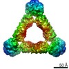 22830MC M: map data used to model this data C: citing same article ( |
|---|---|
| Similar structure data |
-Links
 PDBj
PDBj


- Assembly
| Deposited unit | 
| |||||||||||||||||||||||||||||||||||||||||||||||||||||||||||||||||||||||||||||||||||||||||||||||||||||||||||||||||||||||||||||||||||||||||||||
|---|---|---|---|---|---|---|---|---|---|---|---|---|---|---|---|---|---|---|---|---|---|---|---|---|---|---|---|---|---|---|---|---|---|---|---|---|---|---|---|---|---|---|---|---|---|---|---|---|---|---|---|---|---|---|---|---|---|---|---|---|---|---|---|---|---|---|---|---|---|---|---|---|---|---|---|---|---|---|---|---|---|---|---|---|---|---|---|---|---|---|---|---|---|---|---|---|---|---|---|---|---|---|---|---|---|---|---|---|---|---|---|---|---|---|---|---|---|---|---|---|---|---|---|---|---|---|---|---|---|---|---|---|---|---|---|---|---|---|---|---|---|---|
| 1 |
| |||||||||||||||||||||||||||||||||||||||||||||||||||||||||||||||||||||||||||||||||||||||||||||||||||||||||||||||||||||||||||||||||||||||||||||
| Noncrystallographic symmetry (NCS) | NCS domain:
NCS domain segments:
|