+ Open data
- Basic information
| Entry | Database: PDB / ID: 7k9l | |||||||||||||||||||||||||||
|---|---|---|---|---|---|---|---|---|---|---|---|---|---|---|---|---|---|---|---|---|---|---|---|---|---|---|---|---|
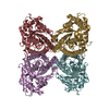
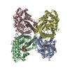
| Title | Aldolase, rabbit muscle (no beam-tilt refinement) | |||||||||||||||||||||||||||
 Components Components | Fructose-bisphosphate aldolase A | |||||||||||||||||||||||||||
 Keywords Keywords | LYASE / glycolysis / gluconeogenesis / Carbohydrate degradation / Homotetramer | |||||||||||||||||||||||||||
| Function / homology |  Function and homology information Function and homology informationnegative regulation of Arp2/3 complex-mediated actin nucleation / fructose-bisphosphate aldolase / fructose-bisphosphate aldolase activity / M band / I band / glycolytic process / protein homotetramerization / positive regulation of cell migration Similarity search - Function | |||||||||||||||||||||||||||
| Biological species |  | |||||||||||||||||||||||||||
| Method | ELECTRON MICROSCOPY / single particle reconstruction / cryo EM / Resolution: 4.9 Å | |||||||||||||||||||||||||||
 Authors Authors | Cianfrocco, M.A. / Kearns, S.E. / Cash, J.N. / Li, Y. | |||||||||||||||||||||||||||
| Funding support |  United States, 2items United States, 2items
| |||||||||||||||||||||||||||
 Citation Citation |  Journal: IUCrJ / Year: 2020 Journal: IUCrJ / Year: 2020Title: High-resolution cryo-EM using beam-image shift at 200 keV. Authors: Jennifer N Cash / Sarah Kearns / Yilai Li / Michael A Cianfrocco /  Abstract: Recent advances in single-particle cryo-electron microscopy (cryo-EM) data collection utilize beam-image shift to improve throughput. Despite implementation on 300 keV cryo-EM instruments, it ...Recent advances in single-particle cryo-electron microscopy (cryo-EM) data collection utilize beam-image shift to improve throughput. Despite implementation on 300 keV cryo-EM instruments, it remains unknown how well beam-image-shift data collection affects data quality on 200 keV instruments and the extent to which aberrations can be computationally corrected. To test this, a cryo-EM data set for aldolase was collected at 200 keV using beam-image shift and analyzed. This analysis shows that the instrument beam tilt and particle motion initially limited the resolution to 4.9 Å. After particle polishing and iterative rounds of aberration correction in , a 2.8 Å resolution structure could be obtained. This analysis demonstrates that software correction of microscope aberrations can provide a significant improvement in resolution at 200 keV. | |||||||||||||||||||||||||||
| History |
|
- Structure visualization
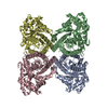
| Movie |
 Movie viewer Movie viewer |
|---|---|
| Structure viewer | Molecule:  Molmil Molmil Jmol/JSmol Jmol/JSmol |
- Downloads & links
- Download
| PDBx/mmCIF format |  7k9l.cif.gz 7k9l.cif.gz | 232.2 KB | Display |  PDBx/mmCIF format PDBx/mmCIF format |
|---|---|---|---|---|
| PDB format |  pdb7k9l.ent.gz pdb7k9l.ent.gz | 189.7 KB | Display |  PDB format PDB format |
| PDBx/mmJSON format |  7k9l.json.gz 7k9l.json.gz | Tree view |  PDBx/mmJSON format PDBx/mmJSON format | |
| Others |  Other downloads Other downloads |
-Validation report
| Summary document |  7k9l_validation.pdf.gz 7k9l_validation.pdf.gz | 971.1 KB | Display |  wwPDB validaton report wwPDB validaton report |
|---|---|---|---|---|
| Full document |  7k9l_full_validation.pdf.gz 7k9l_full_validation.pdf.gz | 989.7 KB | Display | |
| Data in XML |  7k9l_validation.xml.gz 7k9l_validation.xml.gz | 46.5 KB | Display | |
| Data in CIF |  7k9l_validation.cif.gz 7k9l_validation.cif.gz | 71.3 KB | Display | |
| Arichive directory |  https://data.pdbj.org/pub/pdb/validation_reports/k9/7k9l https://data.pdbj.org/pub/pdb/validation_reports/k9/7k9l ftp://data.pdbj.org/pub/pdb/validation_reports/k9/7k9l ftp://data.pdbj.org/pub/pdb/validation_reports/k9/7k9l | HTTPS FTP |
-Related structure data
| Related structure data |  22754MC  7k9xC  7ka2C  7ka3C  7ka4C M: map data used to model this data C: citing same article ( |
|---|---|
| Similar structure data | |
| EM raw data |  EMPIAR-10519 (Title: Single particle cryo electron microscopy of aldolase (rabbit, muscle) using beam-tilt on Talos Arctica EMPIAR-10519 (Title: Single particle cryo electron microscopy of aldolase (rabbit, muscle) using beam-tilt on Talos ArcticaData size: 87.6 Data #1: Unaligned micrographs of aldolase collected with beam-tilt at 200 kV [micrographs - single frame]) |
- Links
Links
- Assembly
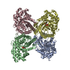
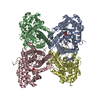
| Deposited unit | 
|
|---|---|
| 1 |
|
- Components
| #1: Protein | Mass: 39263.672 Da / Num. of mol.: 4 Source method: isolated from a genetically manipulated source Source: (gene. exp.)   Has protein modification | N | |
|---|
-Experimental details
-Experiment
| Experiment | Method: ELECTRON MICROSCOPY |
|---|---|
| EM experiment | Aggregation state: PARTICLE / 3D reconstruction method: single particle reconstruction |
- Sample preparation
| Component | Name: Homotetramer of aldolase / Type: COMPLEX / Entity ID: all / Source: NATURAL | ||||||||||||
|---|---|---|---|---|---|---|---|---|---|---|---|---|---|
| Molecular weight | Value: 157 kDa/nm / Experimental value: YES | ||||||||||||
| Source (natural) | Organism:  | ||||||||||||
| Buffer solution | pH: 7.5 | ||||||||||||
| Buffer component |
| ||||||||||||
| Specimen | Conc.: 1.6 mg/ml / Embedding applied: NO / Shadowing applied: NO / Staining applied: NO / Vitrification applied: YES Details: Pure aldolase isolated from rabbit muscle was purchased as a lyophilized powder (Sigma Aldrich) and solubilized in 20 mM HEPES (pH 7.5), 50 mM NaCl at 1.6 mg/ml. Sample was blotted for 4 ...Details: Pure aldolase isolated from rabbit muscle was purchased as a lyophilized powder (Sigma Aldrich) and solubilized in 20 mM HEPES (pH 7.5), 50 mM NaCl at 1.6 mg/ml. Sample was blotted for 4 seconds with Whatman No. #1 filter paper immediately prior to plunge freezing in liquid ethane cooled by liquid nitrogen. | ||||||||||||
| Specimen support | Grid material: GOLD / Grid mesh size: 300 divisions/in. / Grid type: UltrAuFoil R1.2/1.3 | ||||||||||||
| Vitrification | Instrument: FEI VITROBOT MARK IV / Cryogen name: ETHANE / Humidity: 95 % / Chamber temperature: 277.15 K |
- Electron microscopy imaging
| Experimental equipment |  Model: Talos Arctica / Image courtesy: FEI Company |
|---|---|
| Microscopy | Model: FEI TALOS ARCTICA |
| Electron gun | Electron source:  FIELD EMISSION GUN / Accelerating voltage: 200 kV / Illumination mode: OTHER FIELD EMISSION GUN / Accelerating voltage: 200 kV / Illumination mode: OTHER |
| Electron lens | Mode: BRIGHT FIELD / Nominal magnification: 45000 X / Nominal defocus max: 2 nm / Nominal defocus min: 0.8 nm / Cs: 2.7 mm / C2 aperture diameter: 100 µm |
| Image recording | Average exposure time: 10 sec. / Electron dose: 42 e/Å2 / Film or detector model: GATAN K2 BASE (4k x 4k) Details: Images were collected on a Talos Arctica transmission electron microscope (Thermo Fisher) operating at 200 keV with a gun lens of 6, a spot size of 6, 70 um C2 aperture and 100 um objective ...Details: Images were collected on a Talos Arctica transmission electron microscope (Thermo Fisher) operating at 200 keV with a gun lens of 6, a spot size of 6, 70 um C2 aperture and 100 um objective aperture using beam-image shift. Movies were collected using a K2 direct electron detector (Gatan Inc.) operating in counting mode at 45,000x corresponding to a physical pixel size of 0.91 A/pixel with a 10 sec exposure using 200 ms per frame. Using an exposure rate of 4.204 e/pix/sec, each movie had a total dose of approximately 42 e/A2 for the 2,111 movies over a defocus 0.8-2 um. |
- Processing
| Software | Name: PHENIX / Version: 1.14_3260: / Classification: refinement | ||||||||||||||||
|---|---|---|---|---|---|---|---|---|---|---|---|---|---|---|---|---|---|
| EM software |
| ||||||||||||||||
| CTF correction | Type: PHASE FLIPPING AND AMPLITUDE CORRECTION | ||||||||||||||||
| Particle selection | Num. of particles selected: 718578 | ||||||||||||||||
| 3D reconstruction | Resolution: 4.9 Å / Resolution method: FSC 0.143 CUT-OFF / Num. of particles: 186471 / Symmetry type: POINT |
 Movie
Movie Controller






 PDBj
PDBj
