[English] 日本語
Yorodumi
- PDB-7jy8: Analysis of a strand exchange reaction with a mini filament of 9-... -

+
Open data


ID or keywords:

Loading...

-
Basic information

Entry
Database: PDB / ID: 7jy8
TitleAnalysis of a strand exchange reaction with a mini filament of 9-RecA, 27-mer ssDNA, partially-homologous 67 bp dsDNA and ATPgammaS
Components
  • DNA (27-MER)
  • DNA (5'-D(P*AP*AP*AP*AP*AP*AP*AP*AP*AP*A)-3')
  • Protein RecA
KeywordsDNA BINDING PROTEIN/DNA / Recombination / DNA repair / DNA BINDING PROTEIN / DNA BINDING PROTEIN-DNA complex
Function / homology
Function and homology information


DNA polymerase V complex / homologous recombination / SOS response / recombinational repair / ATP-dependent DNA damage sensor activity / response to ionizing radiation / translesion synthesis / ATP-dependent activity, acting on DNA / cell motility / single-stranded DNA binding ...DNA polymerase V complex / homologous recombination / SOS response / recombinational repair / ATP-dependent DNA damage sensor activity / response to ionizing radiation / translesion synthesis / ATP-dependent activity, acting on DNA / cell motility / single-stranded DNA binding / DNA-binding transcription factor binding / DNA recombination / damaged DNA binding / DNA repair / DNA damage response / ATP hydrolysis activity / ATP binding / cytosol / cytoplasm
Similarity search - Function
DNA recombination/repair protein RecA, conserved site / DNA recombination and repair protein RecA, C-terminal / : / RecA C-terminal domain / recA signature. / DNA recombination and repair protein RecA / : / recA bacterial DNA recombination protein / DNA recombination and repair protein RecA, monomer-monomer interface / RecA family profile 2. ...DNA recombination/repair protein RecA, conserved site / DNA recombination and repair protein RecA, C-terminal / : / RecA C-terminal domain / recA signature. / DNA recombination and repair protein RecA / : / recA bacterial DNA recombination protein / DNA recombination and repair protein RecA, monomer-monomer interface / RecA family profile 2. / DNA recombination and repair protein RecA-like, ATP-binding domain / RecA family profile 1. / ATPases associated with a variety of cellular activities / AAA+ ATPase domain / P-loop containing nucleoside triphosphate hydrolase
Similarity search - Domain/homology
PHOSPHOTHIOPHOSPHORIC ACID-ADENYLATE ESTER / DNA / DNA (> 10) / Protein RecA / Protein RecA
Similarity search - Component
Biological speciesEscherichia coli (E. coli)
MethodELECTRON MICROSCOPY / single particle reconstruction / cryo EM / Resolution: 2.4 Å
AuthorsPavletich, N.P.
Funding support United States, 1items
OrganizationGrant numberCountry
Howard Hughes Medical Institute (HHMI) United States
CitationJournal: Nature / Year: 2020
Title: Mechanism of strand exchange from RecA-DNA synaptic and D-loop structures.
Authors: Haijuan Yang / Chun Zhou / Ankita Dhar / Nikola P Pavletich /
Abstract: The strand-exchange reaction is central to homologous recombination. It is catalysed by the RecA family of ATPases, which form a helical filament with single-stranded DNA (ssDNA) and ATP. This ...The strand-exchange reaction is central to homologous recombination. It is catalysed by the RecA family of ATPases, which form a helical filament with single-stranded DNA (ssDNA) and ATP. This filament binds to a donor double-stranded DNA (dsDNA) to form synaptic filaments, which search for homology and then catalyse the exchange of the complementary strand, forming either a new heteroduplex or-if homology is limited-a D-loop. How synaptic filaments form, search for homology and catalyse strand exchange is poorly understood. Here we report the cryo-electron microscopy analysis of synaptic mini-filaments with both non-complementary and partially complementary dsDNA, and structures of RecA-D-loop complexes containing a 10- or a 12-base-pair heteroduplex. The C-terminal domain of RecA binds to dsDNA and directs it to the RecA L2 loop, which inserts into and opens up the duplex. The opening propagates through RecA sequestering the homologous strand at a secondary DNA-binding site, which frees the complementary strand to sample pairing with the ssDNA. At each RecA step, there is a roughly 20% probability that duplex opening will terminate and the as-yet-unopened dsDNA portion will bind to another C-terminal domain. Homology suppresses this process, through the cooperation of heteroduplex pairing with the binding of ssDNA to the secondary site, to extend dsDNA opening. This mechanism locally limits the length of ssDNA sampled for pairing if homology is not encountered, and could allow for the formation of multiple, widely separated synapses on the donor dsDNA, which would increase the likelihood of encountering homology. These findings provide key mechanistic insights into homologous recombination.
History
DepositionAug 29, 2020Deposition site: RCSB / Processing site: RCSB
Revision 1.0Nov 4, 2020Provider: repository / Type: Initial release
Revision 1.1Nov 11, 2020Group: Database references / Category: citation / citation_author
Item: _citation.journal_volume / _citation.page_first ..._citation.journal_volume / _citation.page_first / _citation.page_last / _citation_author.identifier_ORCID
Revision 1.2Mar 6, 2024Group: Data collection / Database references / Refinement description
Category: chem_comp_atom / chem_comp_bond ...chem_comp_atom / chem_comp_bond / database_2 / em_3d_fitting_list / pdbx_initial_refinement_model / refine
Item: _database_2.pdbx_DOI / _database_2.pdbx_database_accession ..._database_2.pdbx_DOI / _database_2.pdbx_database_accession / _em_3d_fitting_list.accession_code / _em_3d_fitting_list.initial_refinement_model_id / _em_3d_fitting_list.source_name / _em_3d_fitting_list.type / _refine.ls_d_res_high / _refine.ls_d_res_low

-
Structure visualization

Movie
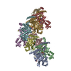
  • Deposited structure unit
  • Imaged by Jmol
  • Download
  • Superimposition on EM map
  • EMDB-22524
  • Imaged by UCSF Chimera
  • Download
Movie viewer
Structure viewerMolecule:
MolmilJmol/JSmol

Downloads & links

-
Assembly

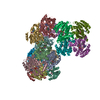
Deposited unit
A: Protein RecA
B: Protein RecA
C: Protein RecA
D: Protein RecA
E: Protein RecA
F: Protein RecA
G: Protein RecA
H: Protein RecA
I: Protein RecA
S: DNA (27-MER)
T: DNA (5'-D(P*AP*AP*AP*AP*AP*AP*AP*AP*AP*A)-3')
hetero molecules


Theoretical massNumber of molelcules
Total (without water)350,78829
Polymers345,86011
Non-polymers4,92818
Water00
1


  • Idetical with deposited unit
  • defined by author
  • Evidence: scanning transmission electron microscopy
TypeNameSymmetry operationNumber
identity operation1_5551

-
Components

#1: Protein
Protein RecA / Recombinase A


Mass: 35960.281 Da / Num. of mol.: 9
Source method: isolated from a genetically manipulated source
Source: (gene. exp.) Escherichia coli (E. coli) / Gene: recA, NCTC11341_01072 / Production host: Escherichia coli (E. coli) / References: UniProt: A0A376NU07, UniProt: P0A7G6*PLUS
#2: DNA chain DNA (27-MER)


Mass: 8168.248 Da / Num. of mol.: 1 / Source method: obtained synthetically / Source: (synth.) Escherichia coli (E. coli)
#3: DNA chain DNA (5'-D(P*AP*AP*AP*AP*AP*AP*AP*AP*AP*A)-3')


Mass: 14049.344 Da / Num. of mol.: 1 / Source method: obtained synthetically / Source: (synth.) Escherichia coli (E. coli)
#4: Chemical
ChemComp-MG / MAGNESIUM ION


Mass: 24.305 Da / Num. of mol.: 9 / Source method: obtained synthetically / Formula: Mg
#5: Chemical
ChemComp-AGS / PHOSPHOTHIOPHOSPHORIC ACID-ADENYLATE ESTER / ATP-GAMMA-S / ADENOSINE 5'-(3-THIOTRIPHOSPHATE) / ADENOSINE 5'-(GAMMA-THIOTRIPHOSPHATE) / ADENOSINE-5'-DIPHOSPHATE MONOTHIOPHOSPHATE


Mass: 523.247 Da / Num. of mol.: 9 / Source method: obtained synthetically / Formula: C10H16N5O12P3S / Comment: ATP-gamma-S, energy-carrying molecule analogue*YM
Has ligand of interestN

-
Experimental details

-
Experiment

ExperimentMethod: ELECTRON MICROSCOPY
EM experimentAggregation state: PARTICLE / 3D reconstruction method: single particle reconstruction

-
Sample preparation

ComponentName: Ensemble of products of a strand exchange reaction with partially-homologous 67 bp dsDNA and ATPgammaS
Type: COMPLEX / Entity ID: #1-#3 / Source: MULTIPLE SOURCES
Molecular weightExperimental value: NO
Source (natural)Organism: Escherichia coli (E. coli)
Buffer solutionpH: 8
SpecimenEmbedding applied: NO / Shadowing applied: NO / Staining applied: NO / Vitrification applied: YES
VitrificationCryogen name: ETHANE

-
Electron microscopy imaging

Experimental equipment
Model: Titan Krios / Image courtesy: FEI Company
MicroscopyModel: TFS KRIOS
Electron gunElectron source: FIELD EMISSION GUN / Accelerating voltage: 300 kV / Illumination mode: FLOOD BEAM
Electron lensMode: BRIGHT FIELD
Image recordingElectron dose: 52 e/Å2 / Film or detector model: GATAN K3 (6k x 4k)

-
Processing

SoftwareName: REFMAC / Version: 5.8.0258 / Classification: refinement
EM software
IDNameCategory
7Omodel fitting
13REFMACmodel refinement
CTF correctionType: PHASE FLIPPING AND AMPLITUDE CORRECTION
3D reconstructionResolution: 2.4 Å / Resolution method: FSC 0.143 CUT-OFF / Num. of particles: 1697726 / Symmetry type: POINT
Atomic model buildingPDB-ID: 3CMW
Accession code: 3CMW / Source name: PDB / Type: experimental model
RefinementResolution: 2.4→2.4 Å / Cor.coef. Fo:Fc: 0.867 / SU B: 11.285 / SU ML: 0.109 / ESU R: 0.189
Stereochemistry target values: MAXIMUM LIKELIHOOD WITH PHASES
Details: HYDROGENS HAVE BEEN ADDED IN THE RIDING POSITIONS
RfactorNum. reflection% reflection
Rwork0.27491 --
obs0.27491 401287 100 %
Solvent computationIon probe radii: 0.8 Å / Shrinkage radii: 0.8 Å / VDW probe radii: 1 Å / Solvent model: MASK
Displacement parametersBiso mean: 29.639 Å2
Baniso -1Baniso -2Baniso -3
1-0.33 Å2-0.16 Å2-0.05 Å2
2---1.31 Å21.46 Å2
3---0.98 Å2
Refinement stepCycle: 1 / Total: 23360
Refine LS restraints
Refine-IDTypeDev idealDev ideal targetNumber
ELECTRON MICROSCOPYr_bond_refined_d0.0060.01323738
ELECTRON MICROSCOPYr_bond_other_d0.0350.01722593
ELECTRON MICROSCOPYr_angle_refined_deg1.5581.63732133
ELECTRON MICROSCOPYr_angle_other_deg2.371.61452506
ELECTRON MICROSCOPYr_dihedral_angle_1_deg6.28352955
ELECTRON MICROSCOPYr_dihedral_angle_2_deg34.47523.591039
ELECTRON MICROSCOPYr_dihedral_angle_3_deg19.172154248
ELECTRON MICROSCOPYr_dihedral_angle_4_deg20.7115124
ELECTRON MICROSCOPYr_chiral_restr0.0650.23184
ELECTRON MICROSCOPYr_gen_planes_refined0.0040.0225800
ELECTRON MICROSCOPYr_gen_planes_other0.0050.024413
ELECTRON MICROSCOPYr_nbd_refined
ELECTRON MICROSCOPYr_nbd_other
ELECTRON MICROSCOPYr_nbtor_refined
ELECTRON MICROSCOPYr_nbtor_other
ELECTRON MICROSCOPYr_xyhbond_nbd_refined
ELECTRON MICROSCOPYr_xyhbond_nbd_other
ELECTRON MICROSCOPYr_metal_ion_refined
ELECTRON MICROSCOPYr_metal_ion_other
ELECTRON MICROSCOPYr_symmetry_vdw_refined
ELECTRON MICROSCOPYr_symmetry_vdw_other
ELECTRON MICROSCOPYr_symmetry_hbond_refined
ELECTRON MICROSCOPYr_symmetry_hbond_other
ELECTRON MICROSCOPYr_symmetry_metal_ion_refined
ELECTRON MICROSCOPYr_symmetry_metal_ion_other
ELECTRON MICROSCOPYr_mcbond_it3.3584.53411847
ELECTRON MICROSCOPYr_mcbond_other3.3574.53311846
ELECTRON MICROSCOPYr_mcangle_it6.0522.5914793
ELECTRON MICROSCOPYr_mcangle_other6.0522.59414794
ELECTRON MICROSCOPYr_scbond_it3.4865.08611891
ELECTRON MICROSCOPYr_scbond_other3.4865.08511892
ELECTRON MICROSCOPYr_scangle_it
ELECTRON MICROSCOPYr_scangle_other6.49324.84117341
ELECTRON MICROSCOPYr_long_range_B_refined10.58765.43923392
ELECTRON MICROSCOPYr_long_range_B_other10.58765.43723393
ELECTRON MICROSCOPYr_rigid_bond_restr
ELECTRON MICROSCOPYr_sphericity_free
ELECTRON MICROSCOPYr_sphericity_bonded
LS refinement shellResolution: 2.5→2.565 Å / Total num. of bins used: 20
RfactorNum. reflection% reflection
Rfree0 0 -
Rwork0.55 29628 -
obs--100 %
Refinement TLS params.

Method: refined / Refine-ID: ELECTRON MICROSCOPY

IDL112)L122)L132)L222)L232)L332)S11 (Å °)S12 (Å °)S13 (Å °)S21 (Å °)S22 (Å °)S23 (Å °)S31 (Å °)S32 (Å °)S33 (Å °)T112)T122)T132)T222)T232)T332)Origin x (Å)Origin y (Å)Origin z (Å)
11.37340.1803-0.61761.8241-1.19231.4679-0.11320.07420.1426-0.0968-0.0913-0.184-0.09320.0950.20450.2106-0.15920.0510.2707-0.04730.26958.747384.058242.2809
20.4132-0.27760.03061.2317-0.39540.14580.1073-0.03910.3536-0.17960.13520.45190.041-0.1169-0.24250.3066-0.0718-0.00580.57450.11140.770938.39996.872124.2938
30.54910.06950.43411.6024-0.88620.902-0.06880.01580.06040.03350.05440.0811-0.09820.00010.01440.2171-0.00920.00620.3026-0.07270.247143.918165.248358.5618
41.86860.09462.29295.6234-2.115.02360.21180.20680.263-0.168-0.17541.40780.5202-0.2748-0.03640.2963-0.163-0.14990.3492-0.03260.521723.715952.682637.5031
51.8921-0.32631.04541.53010.04392.0119-0.0092-0.101-0.067-0.03810.01720.2019-0.0217-0.2218-0.00790.0990.00990.00030.267-0.07230.163551.829141.342474.0012
63.7348-3.28163.38866.51341.16957.92630.19640.2909-0.9281-0.14010.05871.21550.33710.5281-0.25510.35150.0362-0.06530.1969-0.23250.535153.066917.076553.5987
71.42780.38740.71821.04851.07151.51350.1135-0.0223-0.19340.1329-0.07320.02630.1158-0.0854-0.04040.1927-0.02240.00330.296-0.0720.192675.710836.341990.8199
83.83281.38581.94133.33493.70876.66460.24360.1601-1.1287-0.3007-0.0157-0.61640.2020.5867-0.22790.16610.11620.08110.5187-0.13860.61697.973624.333571.8392
91.2327-0.0451-0.31311.62381.25711.5934-0.064-0.0296-0.06740.17610.1377-0.1810.09350.0678-0.07380.21050.0078-0.00470.267-0.06640.151692.163754.6641107.8818
102.76370.0646-2.29884.91022.97115.20230.18810.3531-0.1196-0.02970.0853-1.117-0.33130.3219-0.27340.2569-0.17590.13510.3565-0.01060.4531114.259667.030989.8401
112.9408-0.3514-1.3971.57770.15852.14780.0005-0.12940.0517-0.07160.0172-0.20960.03780.3179-0.01770.08060.07060.00520.2804-0.05890.10983.525978.8066123.8328
124.1254-0.856-2.95876.8703-1.90553.1210.21180.16181.2574-0.07150.3806-1.0207-0.3188-0.2027-0.59240.7276-0.03230.15540.40490.3130.751285.5351103.6201105.1088
132.53320.2347-0.8911.7511-0.59512.09710.1088-0.18370.22820.1357-0.0660.0471-0.15010.0513-0.04270.0610.0281-0.00780.2478-0.08580.122557.584784.456138.2234
147.51362.55-1.54924.8175-5.85787.5878-0.03520.1691.5269-0.28250.36760.53730.2544-0.6402-0.33240.23120.2218-0.0860.52940.1060.458138.544898.2863117.9144
151.4010.5484-0.02991.0908-0.58771.31630.0142-0.1149-0.05580.13150.03610.16190.0578-0.042-0.05040.1495-0.00240.00180.3237-0.05340.217938.462865.7762152.2377
161.81490.76321.96657.3285-3.36795.02530.20120.13430.2564-0.28660.01271.16840.4913-0.2448-0.21390.2387-0.1232-0.18340.5006-0.06540.410917.858655.96131.1223
170.52590.57240.30622.9899-0.51161.50890.1061-0.1106-0.3330.102-0.0571-0.15530.0644-0.0022-0.0490.13450.0787-0.09540.2816-0.02290.327843.59839.9852167.4269
187.2716-0.81233.23814.361-2.48535.48510.31140.1436-1.0519-0.69990.32820.91951.23870.1915-0.63960.57230.2171-0.32080.3017-0.22160.643342.302316.8563146.9072
Refinement TLS group
IDRefine-IDRefine TLS-IDAuth asym-IDAuth seq-ID
1ELECTRON MICROSCOPY1A37 - 269
2ELECTRON MICROSCOPY1A500 - 502
3ELECTRON MICROSCOPY1S24 - 27
4ELECTRON MICROSCOPY2A270 - 333
5ELECTRON MICROSCOPY3A1 - 36
6ELECTRON MICROSCOPY3B37 - 269
7ELECTRON MICROSCOPY3B500 - 502
8ELECTRON MICROSCOPY3S21 - 23
9ELECTRON MICROSCOPY3T30 - 33
10ELECTRON MICROSCOPY4B270 - 333
11ELECTRON MICROSCOPY5B1 - 36
12ELECTRON MICROSCOPY5C37 - 269
13ELECTRON MICROSCOPY5C500 - 502
14ELECTRON MICROSCOPY5S18 - 20
15ELECTRON MICROSCOPY5T34 - 36
16ELECTRON MICROSCOPY6C270 - 333
17ELECTRON MICROSCOPY7C1 - 36
18ELECTRON MICROSCOPY7D37 - 269
19ELECTRON MICROSCOPY7D500 - 502
20ELECTRON MICROSCOPY7S15 - 17
21ELECTRON MICROSCOPY7T37 - 39
22ELECTRON MICROSCOPY8D270 - 333
23ELECTRON MICROSCOPY9D1 - 36
24ELECTRON MICROSCOPY9E37 - 269
25ELECTRON MICROSCOPY9E500 - 502
26ELECTRON MICROSCOPY9S12 - 14
27ELECTRON MICROSCOPY10E270 - 333
28ELECTRON MICROSCOPY11E1 - 36
29ELECTRON MICROSCOPY11F37 - 269
30ELECTRON MICROSCOPY11F500 - 502
31ELECTRON MICROSCOPY11S9 - 11
32ELECTRON MICROSCOPY12F270 - 333
33ELECTRON MICROSCOPY13F1 - 36
34ELECTRON MICROSCOPY13G37 - 269
35ELECTRON MICROSCOPY13G500 - 502
36ELECTRON MICROSCOPY13S6 - 8
37ELECTRON MICROSCOPY14G270 - 333
38ELECTRON MICROSCOPY15G1 - 36
39ELECTRON MICROSCOPY15H37 - 269
40ELECTRON MICROSCOPY15H500 - 502
41ELECTRON MICROSCOPY15S3 - 5
42ELECTRON MICROSCOPY16H270 - 333
43ELECTRON MICROSCOPY17H1 - 36
44ELECTRON MICROSCOPY17I34 - 269
45ELECTRON MICROSCOPY17I500 - 502
46ELECTRON MICROSCOPY17S1 - 2
47ELECTRON MICROSCOPY18I270 - 333

+
About Yorodumi

-
News

-
Feb 9, 2022. New format data for meta-information of EMDB entries

New format data for meta-information of EMDB entries

  • Version 3 of the EMDB header file is now the official format.
  • The previous official version 1.9 will be removed from the archive.

Related info.:EMDB header

External links:wwPDB to switch to version 3 of the EMDB data model

-
Aug 12, 2020. Covid-19 info

Covid-19 info

URL: https://pdbj.org/emnavi/covid19.php

New page: Covid-19 featured information page in EM Navigator.

Related info.:Covid-19 info / Mar 5, 2020. Novel coronavirus structure data

+
Mar 5, 2020. Novel coronavirus structure data

Novel coronavirus structure data

Related info.:Yorodumi Speices / Aug 12, 2020. Covid-19 info

External links:COVID-19 featured content - PDBj / Molecule of the Month (242):Coronavirus Proteases

+
Jan 31, 2019. EMDB accession codes are about to change! (news from PDBe EMDB page)

EMDB accession codes are about to change! (news from PDBe EMDB page)

  • The allocation of 4 digits for EMDB accession codes will soon come to an end. Whilst these codes will remain in use, new EMDB accession codes will include an additional digit and will expand incrementally as the available range of codes is exhausted. The current 4-digit format prefixed with “EMD-” (i.e. EMD-XXXX) will advance to a 5-digit format (i.e. EMD-XXXXX), and so on. It is currently estimated that the 4-digit codes will be depleted around Spring 2019, at which point the 5-digit format will come into force.
  • The EM Navigator/Yorodumi systems omit the EMD- prefix.

Related info.:Q: What is EMD? / ID/Accession-code notation in Yorodumi/EM Navigator

External links:EMDB Accession Codes are Changing Soon! / Contact to PDBj

+
Jul 12, 2017. Major update of PDB

Major update of PDB

  • wwPDB released updated PDB data conforming to the new PDBx/mmCIF dictionary.
  • This is a major update changing the version number from 4 to 5, and with Remediation, in which all the entries are updated.
  • In this update, many items about electron microscopy experimental information are reorganized (e.g. em_software).
  • Now, EM Navigator and Yorodumi are based on the updated data.

External links:wwPDB Remediation / Enriched Model Files Conforming to OneDep Data Standards Now Available in the PDB FTP Archive

-
Yorodumi

Thousand views of thousand structures

  • Yorodumi is a browser for structure data from EMDB, PDB, SASBDB, etc.
  • This page is also the successor to EM Navigator detail page, and also detail information page/front-end page for Omokage search.
  • The word "yorodu" (or yorozu) is an old Japanese word meaning "ten thousand". "mi" (miru) is to see.

Related info.:EMDB / PDB / SASBDB / Comparison of 3 databanks / Yorodumi Search / Aug 31, 2016. New EM Navigator & Yorodumi / Yorodumi Papers / Jmol/JSmol / Function and homology information / Changes in new EM Navigator and Yorodumi

Read more