Chloride channel ClC-7 / : / Chloride channel, voltage gated / Chloride channel, core / Voltage gated chloride channel / Domain in cystathionine beta-synthase and other proteins. / CBS domain superfamily / CBS domain / CBS domain / CBS domain profile. Similarity search - Domain/homology
Chem-0J1 / ADENOSINE-5'-TRIPHOSPHATE / Chloride channel protein / Chloride channel protein Similarity search - Component
Biological species
Gallus gallus (chicken)
Method
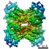
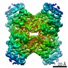
ELECTRON MICROSCOPY / single particle reconstruction / cryo EM / Resolution: 2.92 Å
National Institutes of Health/National Institute of General Medical Sciences (NIH/NIGMS)
GM103310
United States
National Institutes of Health/National Cancer Institute (NIH/NCI)
P30 CA008748
United States
Citation
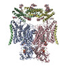
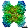
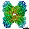
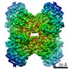
Journal: Elife / Year: 2020 Title: Cryo-EM structure of the lysosomal chloride-proton exchanger CLC-7 in complex with OSTM1. Authors: Marina Schrecker / Julia Korobenko / Richard K Hite / Abstract: The chloride-proton exchanger CLC-7 plays critical roles in lysosomal homeostasis and bone regeneration and its mutation can lead to osteopetrosis, lysosomal storage disease and neurological ...The chloride-proton exchanger CLC-7 plays critical roles in lysosomal homeostasis and bone regeneration and its mutation can lead to osteopetrosis, lysosomal storage disease and neurological disorders. In lysosomes and the ruffled border of osteoclasts, CLC-7 requires a β-subunit, OSTM1, for stability and activity. Here, we present electron cryomicroscopy structures of CLC-7 in occluded states by itself and in complex with OSTM1, determined at resolutions up to 2.8 Å. In the complex, the luminal surface of CLC-7 is entirely covered by a dimer of the heavily glycosylated and disulfide-bonded OSTM1, which serves to protect CLC-7 from the degradative environment of the lysosomal lumen. OSTM1 binding does not induce large-scale rearrangements of CLC-7, but does have minor effects on the conformation of the ion-conduction pathway, potentially contributing to its regulatory role. These studies provide insights into the role of OSTM1 and serve as a foundation for understanding the mechanisms of CLC-7 regulation.
In the structure databanks used in Yorodumi, some data are registered as the other names, "COVID-19 virus" and "2019-nCoV". Here are the details of the virus and the list of structure data.
Jan 31, 2019. EMDB accession codes are about to change! (news from PDBe EMDB page)
EMDB accession codes are about to change! (news from PDBe EMDB page)
The allocation of 4 digits for EMDB accession codes will soon come to an end. Whilst these codes will remain in use, new EMDB accession codes will include an additional digit and will expand incrementally as the available range of codes is exhausted. The current 4-digit format prefixed with “EMD-” (i.e. EMD-XXXX) will advance to a 5-digit format (i.e. EMD-XXXXX), and so on. It is currently estimated that the 4-digit codes will be depleted around Spring 2019, at which point the 5-digit format will come into force.
The EM Navigator/Yorodumi systems omit the EMD- prefix.
Related info.:Q: What is EMD? / ID/Accession-code notation in Yorodumi/EM Navigator
Yorodumi is a browser for structure data from EMDB, PDB, SASBDB, etc.
This page is also the successor to EM Navigator detail page, and also detail information page/front-end page for Omokage search.
The word "yorodu" (or yorozu) is an old Japanese word meaning "ten thousand". "mi" (miru) is to see.
Related info.:EMDB / PDB / SASBDB / Comparison of 3 databanks / Yorodumi Search / Aug 31, 2016. New EM Navigator & Yorodumi / Yorodumi Papers / Jmol/JSmol / Function and homology information / Changes in new EM Navigator and Yorodumi
 Movie
Movie Controller


 Open data
 Basic information
 Components
 Keywords
 Function and homology information
 Authors
 United States, 2items
  Citation
 Structure visualization
 Downloads & links
 Other downloads


 PDBj
PDBj
 Assembly
 Homo sapiens (human) / References: UniProt: Q5ZL60, UniProt: A0A1D5PS57*PLUS


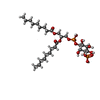

 Mass: 507.181 Da / Num. of mol.: 2 / Source method: obtained synthetically / Formula: C10H16N5O13P3 / Feature type: SUBJECT OF INVESTIGATION / Comment: ATP, energy-carrying molecule*YM
 Mass: 24.305 Da / Num. of mol.: 2 / Source method: obtained synthetically / Formula: Mg
 Mass: 35.453 Da / Num. of mol.: 6 / Source method: obtained synthetically / Formula: Cl
 Mass: 694.640 Da / Num. of mol.: 2 / Source method: obtained synthetically / Formula: C27H52O16P2 / Feature type: SUBJECT OF INVESTIGATION
 Sample preparation
 Electron microscopy imaging
 FIELD EMISSION GUN / Accelerating voltage: 300 kV / Illumination mode: FLOOD BEAM
 Processing