[English] 日本語
 Yorodumi
- PDB-7fg2: Minor cryo-EM structure of S protein trimer of SARS-CoV2 with K-8... -
+ Open data
- Basic information
| Entry | Database: PDB / ID: 7fg2 | |||||||||
|---|---|---|---|---|---|---|---|---|---|---|
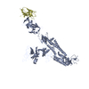
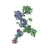
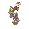
| Title | Minor cryo-EM structure of S protein trimer of SARS-CoV2 with K-874A VHH, composite map | |||||||||
 Components Components |
| |||||||||
 Keywords Keywords | PROTEIN BINDING / Antibody | |||||||||
| Function / homology |  Function and homology information Function and homology informationsymbiont-mediated disruption of host tissue / Maturation of spike protein / Translation of Structural Proteins / Virion Assembly and Release / host cell surface / host extracellular space / viral translation / symbiont-mediated-mediated suppression of host tetherin activity / Induction of Cell-Cell Fusion / structural constituent of virion ...symbiont-mediated disruption of host tissue / Maturation of spike protein / Translation of Structural Proteins / Virion Assembly and Release / host cell surface / host extracellular space / viral translation / symbiont-mediated-mediated suppression of host tetherin activity / Induction of Cell-Cell Fusion / structural constituent of virion / membrane fusion / entry receptor-mediated virion attachment to host cell / Attachment and Entry / host cell endoplasmic reticulum-Golgi intermediate compartment membrane / positive regulation of viral entry into host cell / receptor-mediated virion attachment to host cell / host cell surface receptor binding / symbiont-mediated suppression of host innate immune response / receptor ligand activity / endocytosis involved in viral entry into host cell / fusion of virus membrane with host plasma membrane / fusion of virus membrane with host endosome membrane / viral envelope / symbiont entry into host cell / virion attachment to host cell / SARS-CoV-2 activates/modulates innate and adaptive immune responses / host cell plasma membrane / virion membrane / identical protein binding / membrane / plasma membrane Similarity search - Function | |||||||||
| Biological species |   Homo sapiens (human) Homo sapiens (human) | |||||||||
| Method | ELECTRON MICROSCOPY / single particle reconstruction / cryo EM / Resolution: 4.4 Å | |||||||||
 Authors Authors | Song, C. / Murata, K. / Katayama, K. | |||||||||
| Funding support |  Japan, 2items Japan, 2items
| |||||||||
 Citation Citation |  Journal: PLoS Pathog / Year: 2021 Journal: PLoS Pathog / Year: 2021Title: Nasal delivery of single-domain antibody improves symptoms of SARS-CoV-2 infection in an animal model. Authors: Kei Haga / Reiko Takai-Todaka / Yuta Matsumura / Chihong Song / Tomomi Takano / Takuto Tojo / Atsushi Nagami / Yuki Ishida / Hidekazu Masaki / Masayuki Tsuchiya / Toshiki Ebisudani / Shinya ...Authors: Kei Haga / Reiko Takai-Todaka / Yuta Matsumura / Chihong Song / Tomomi Takano / Takuto Tojo / Atsushi Nagami / Yuki Ishida / Hidekazu Masaki / Masayuki Tsuchiya / Toshiki Ebisudani / Shinya Sugimoto / Toshiro Sato / Hiroyuki Yasuda / Koichi Fukunaga / Akihito Sawada / Naoto Nemoto / Kazuyoshi Murata / Takuya Morimoto / Kazuhiko Katayama /  Abstract: The severe acute respiratory syndrome coronavirus 2 (SARS-CoV-2) that causes the disease COVID-19 can lead to serious symptoms, such as severe pneumonia, in the elderly and those with underlying ...The severe acute respiratory syndrome coronavirus 2 (SARS-CoV-2) that causes the disease COVID-19 can lead to serious symptoms, such as severe pneumonia, in the elderly and those with underlying medical conditions. While vaccines are now available, they do not work for everyone and therapeutic drugs are still needed, particularly for treating life-threatening conditions. Here, we showed nasal delivery of a new, unmodified camelid single-domain antibody (VHH), termed K-874A, effectively inhibited SARS-CoV-2 titers in infected lungs of Syrian hamsters without causing weight loss and cytokine induction. In vitro studies demonstrated that K-874A neutralized SARS-CoV-2 in both VeroE6/TMPRSS2 and human lung-derived alveolar organoid cells. Unlike other drug candidates, K-874A blocks viral membrane fusion rather than viral attachment. Cryo-electron microscopy revealed K-874A bound between the receptor binding domain and N-terminal domain of the virus S protein. Further, infected cells treated with K-874A produced fewer virus progeny that were less infective. We propose that direct administration of K-874A to the lung could be a new treatment for preventing the reinfection of amplified virus in COVID-19 patients. | |||||||||
| History |
|
- Structure visualization
| Movie |
 Movie viewer Movie viewer |
|---|---|
| Structure viewer | Molecule:  Molmil Molmil Jmol/JSmol Jmol/JSmol |
- Downloads & links
- Download
| PDBx/mmCIF format |  7fg2.cif.gz 7fg2.cif.gz | 222.2 KB | Display |  PDBx/mmCIF format PDBx/mmCIF format |
|---|---|---|---|---|
| PDB format |  pdb7fg2.ent.gz pdb7fg2.ent.gz | 174.2 KB | Display |  PDB format PDB format |
| PDBx/mmJSON format |  7fg2.json.gz 7fg2.json.gz | Tree view |  PDBx/mmJSON format PDBx/mmJSON format | |
| Others |  Other downloads Other downloads |
-Validation report
| Summary document |  7fg2_validation.pdf.gz 7fg2_validation.pdf.gz | 902.4 KB | Display |  wwPDB validaton report wwPDB validaton report |
|---|---|---|---|---|
| Full document |  7fg2_full_validation.pdf.gz 7fg2_full_validation.pdf.gz | 949.5 KB | Display | |
| Data in XML |  7fg2_validation.xml.gz 7fg2_validation.xml.gz | 43.8 KB | Display | |
| Data in CIF |  7fg2_validation.cif.gz 7fg2_validation.cif.gz | 64.3 KB | Display | |
| Arichive directory |  https://data.pdbj.org/pub/pdb/validation_reports/fg/7fg2 https://data.pdbj.org/pub/pdb/validation_reports/fg/7fg2 ftp://data.pdbj.org/pub/pdb/validation_reports/fg/7fg2 ftp://data.pdbj.org/pub/pdb/validation_reports/fg/7fg2 | HTTPS FTP |
-Related structure data
| Related structure data |  31574MC  7fg3C  7fg7C C: citing same article ( M: map data used to model this data |
|---|---|
| Similar structure data |
- Links
Links
- Assembly
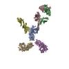
| Deposited unit | 
|
|---|---|
| 1 |
|
- Components
| #1: Protein | Mass: 141297.422 Da / Num. of mol.: 1 Source method: isolated from a genetically manipulated source Source: (gene. exp.)  Gene: S, 2 / Production host:  Homo sapiens (human) / References: UniProt: P0DTC2 Homo sapiens (human) / References: UniProt: P0DTC2 |
|---|---|
| #2: Antibody | Mass: 14362.724 Da / Num. of mol.: 1 Source method: isolated from a genetically manipulated source Source: (gene. exp.)  Homo sapiens (human) / Production host: Homo sapiens (human) / Production host:  Homo sapiens (human) Homo sapiens (human) |
| Has protein modification | Y |
-Experimental details
-Experiment
| Experiment | Method: ELECTRON MICROSCOPY |
|---|---|
| EM experiment | Aggregation state: PARTICLE / 3D reconstruction method: single particle reconstruction |
- Sample preparation
| Component | Name: Minor cryo-EM structure of S protein trimer of SARS-CoV2 with K-874A VHH , composite map Type: COMPLEX / Entity ID: all / Source: RECOMBINANT |
|---|---|
| Molecular weight | Experimental value: NO |
| Source (natural) | Organism:  |
| Source (recombinant) | Organism:  Homo sapiens (human) / Cell: RAW264.7 / Plasmid: cDNA Homo sapiens (human) / Cell: RAW264.7 / Plasmid: cDNA |
| Details of virus | Empty: YES / Enveloped: NO / Isolate: SPECIES / Type: VIRION |
| Natural host | Organism: Mus musculus |
| Virus shell | Diameter: 400 nm / Triangulation number (T number): 3 |
| Buffer solution | pH: 7.4 |
| Specimen | Embedding applied: YES / Shadowing applied: NO / Staining applied: NO / Vitrification applied: YES |
| EM embedding | Material: amorphous ice |
| Vitrification | Cryogen name: ETHANE / Humidity: 95 % / Chamber temperature: 277 K |
- Electron microscopy imaging
| Experimental equipment |  Model: Titan Krios / Image courtesy: FEI Company |
|---|---|
| Microscopy | Model: FEI TITAN KRIOS |
| Electron gun | Electron source:  FIELD EMISSION GUN / Accelerating voltage: 300 kV / Illumination mode: FLOOD BEAM FIELD EMISSION GUN / Accelerating voltage: 300 kV / Illumination mode: FLOOD BEAM |
| Electron lens | Mode: BRIGHT FIELD / Nominal magnification: 64000 X / Nominal defocus max: 2500 nm / Nominal defocus min: 1000 nm / Cs: 0.1 mm / C2 aperture diameter: 100 µm / Alignment procedure: BASIC |
| Specimen holder | Cryogen: NITROGEN / Specimen holder model: FEI TITAN KRIOS AUTOGRID HOLDER / Temperature (max): 77 K / Temperature (min): 76 K |
| Image recording | Average exposure time: 4.6 sec. / Electron dose: 50 e/Å2 / Film or detector model: FEI FALCON III (4k x 4k) / Num. of real images: 6552 |
| Image scans | Width: 4096 / Height: 4096 / Movie frames/image: 32 / Used frames/image: 3-31 |
- Processing
| EM software |
| ||||||||||||||||||||||||||||||||||||
|---|---|---|---|---|---|---|---|---|---|---|---|---|---|---|---|---|---|---|---|---|---|---|---|---|---|---|---|---|---|---|---|---|---|---|---|---|---|
| CTF correction | Type: PHASE FLIPPING ONLY | ||||||||||||||||||||||||||||||||||||
| 3D reconstruction | Resolution: 4.4 Å / Resolution method: FSC 0.143 CUT-OFF / Num. of particles: 51305 / Symmetry type: POINT | ||||||||||||||||||||||||||||||||||||
| Atomic model building | B value: 153 / Protocol: FLEXIBLE FIT / Space: REAL |
 Movie
Movie Controller


 PDBj
PDBj


