Japan Agency for Medical Research and Development (AMED)
JP21am0101071
日本
Japan Society for the Promotion of Science (JSPS)
21K06032
日本
Japan Society for the Promotion of Science (JSPS)
JP18J00191
日本
引用
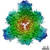
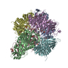
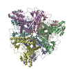
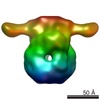
ジャーナル: J Biol Chem / 年: 2021 タイトル: Minimal protein-only RNase P structure reveals insights into tRNA precursor recognition and catalysis. 著者: Takamasa Teramoto / Takeshi Koyasu / Naruhiko Adachi / Masato Kawasaki / Toshio Moriya / Tomoyuki Numata / Toshiya Senda / Yoshimitsu Kakuta / 要旨: Ribonuclease P (RNase P) is an endoribonuclease that catalyzes the processing of the 5' leader sequence of precursor tRNA (pre-tRNA). Ribonucleoprotein RNase P and protein-only RNase P (PRORP) in ...Ribonuclease P (RNase P) is an endoribonuclease that catalyzes the processing of the 5' leader sequence of precursor tRNA (pre-tRNA). Ribonucleoprotein RNase P and protein-only RNase P (PRORP) in eukaryotes have been extensively studied, but the mechanism by which a prokaryotic nuclease recognizes and cleaves pre-tRNA is unclear. To gain insights into this mechanism, we studied homologs of Aquifex RNase P (HARPs), thought to be enzymes of approximately 23 kDa comprising only this nuclease domain. We determined the cryo-EM structure of Aq880, the first identified HARP enzyme. The structure unexpectedly revealed that Aq880 consists of both the nuclease and protruding helical (PrH) domains. Aq880 monomers assemble into a dimer via the PrH domain. Six dimers form a dodecamer with a left-handed one-turn superhelical structure. The structure also revealed that the active site of Aq880 is analogous to that of eukaryotic PRORPs. The pre-tRNA docking model demonstrated that 5' processing of pre-tRNAs is achieved by two adjacent dimers within the dodecamer. One dimer is responsible for catalysis, and the PrH domains of the other dimer are responsible for pre-tRNA elbow recognition. Our study suggests that HARPs measure an invariant distance from the pre-tRNA elbow to cleave the 5' leader sequence, which is analogous to the mechanism of eukaryotic PRORPs and the ribonucleoprotein RNase P. Collectively, these findings shed light on how different types of RNase P enzymes utilize the same pre-tRNA processing.
EMPIAR-11072 (タイトル: Minimal protein-only RNase P structure reveals insights into tRNA precursor recognition and catalysis Data size: 2.0 TB Data #1: Minimal protein-only RNase P structure reveals insights into tRNA precursor recognition and catalysis [micrographs - multiframe])
E: RNA-free ribonuclease P F: RNA-free ribonuclease P C: RNA-free ribonuclease P D: RNA-free ribonuclease P A: RNA-free ribonuclease P B: RNA-free ribonuclease P H: RNA-free ribonuclease P G: RNA-free ribonuclease P J: RNA-free ribonuclease P I: RNA-free ribonuclease P L: RNA-free ribonuclease P K: RNA-free ribonuclease P
 ムービー
ムービー コントローラー


 データを開く
 基本情報
基本情報 要素
要素 キーワード
キーワード 機能・相同性情報
 Aquifex aeolicus (バクテリア)
 データ登録者
 日本, 3件
  引用
引用 構造の表示
構造の表示 ダウンロードとリンク
 その他のダウンロード
 PDBj
PDBj


 集合体
集合体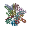
 試料調製
試料調製 電子顕微鏡撮影
 FIELD EMISSION GUN / 加速電圧: 200 kV / 照射モード: FLOOD BEAM
 解析
解析