Japan Agency for Medical Research and Development (AMED)
JP21am0101071
Japan
Japan Society for the Promotion of Science (JSPS)
21K06032
Japan
Japan Society for the Promotion of Science (JSPS)
JP18J00191
Japan
Citation
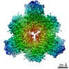
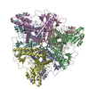
Journal: J Biol Chem / Year: 2021 Title: Minimal protein-only RNase P structure reveals insights into tRNA precursor recognition and catalysis. Authors: Takamasa Teramoto / Takeshi Koyasu / Naruhiko Adachi / Masato Kawasaki / Toshio Moriya / Tomoyuki Numata / Toshiya Senda / Yoshimitsu Kakuta / Abstract: Ribonuclease P (RNase P) is an endoribonuclease that catalyzes the processing of the 5' leader sequence of precursor tRNA (pre-tRNA). Ribonucleoprotein RNase P and protein-only RNase P (PRORP) in ...Ribonuclease P (RNase P) is an endoribonuclease that catalyzes the processing of the 5' leader sequence of precursor tRNA (pre-tRNA). Ribonucleoprotein RNase P and protein-only RNase P (PRORP) in eukaryotes have been extensively studied, but the mechanism by which a prokaryotic nuclease recognizes and cleaves pre-tRNA is unclear. To gain insights into this mechanism, we studied homologs of Aquifex RNase P (HARPs), thought to be enzymes of approximately 23 kDa comprising only this nuclease domain. We determined the cryo-EM structure of Aq880, the first identified HARP enzyme. The structure unexpectedly revealed that Aq880 consists of both the nuclease and protruding helical (PrH) domains. Aq880 monomers assemble into a dimer via the PrH domain. Six dimers form a dodecamer with a left-handed one-turn superhelical structure. The structure also revealed that the active site of Aq880 is analogous to that of eukaryotic PRORPs. The pre-tRNA docking model demonstrated that 5' processing of pre-tRNAs is achieved by two adjacent dimers within the dodecamer. One dimer is responsible for catalysis, and the PrH domains of the other dimer are responsible for pre-tRNA elbow recognition. Our study suggests that HARPs measure an invariant distance from the pre-tRNA elbow to cleave the 5' leader sequence, which is analogous to the mechanism of eukaryotic PRORPs and the ribonucleoprotein RNase P. Collectively, these findings shed light on how different types of RNase P enzymes utilize the same pre-tRNA processing.
EMPIAR-11072 (Title: Minimal protein-only RNase P structure reveals insights into tRNA precursor recognition and catalysis Data size: 2.0 TB Data #1: Minimal protein-only RNase P structure reveals insights into tRNA precursor recognition and catalysis [micrographs - multiframe])
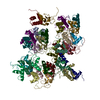
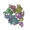
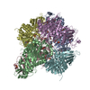
E: RNA-free ribonuclease P F: RNA-free ribonuclease P C: RNA-free ribonuclease P D: RNA-free ribonuclease P A: RNA-free ribonuclease P B: RNA-free ribonuclease P H: RNA-free ribonuclease P G: RNA-free ribonuclease P J: RNA-free ribonuclease P I: RNA-free ribonuclease P L: RNA-free ribonuclease P K: RNA-free ribonuclease P
Conc.: 5 mg/ml / Embedding applied: NO / Shadowing applied: NO / Staining applied: NO / Vitrification applied: YES / Details: This sample was mono-disperse.
Specimen support
Details: The grid was washed by acetone prior to use. / Grid material: COPPER / Grid mesh size: 300 divisions/in. / Grid type: Quantifoil R1.2/1.3
Vitrification
Instrument: FEI VITROBOT MARK IV / Cryogen name: ETHANE / Humidity: 100 % / Chamber temperature: 291 K / Details: Blotting time was 5 seconds (blot force 20)
-
Electron microscopy imaging
Microscopy
Model: TFS TALOS
Electron gun
Electron source: FIELD EMISSION GUN / Accelerating voltage: 200 kV / Illumination mode: FLOOD BEAM
Electron lens
Mode: BRIGHT FIELD / Nominal magnification: 120000 X / Nominal defocus max: 2500 nm / Nominal defocus min: 1000 nm / Cs: 2.7 mm / C2 aperture diameter: 50 µm
Specimen holder
Cryogen: NITROGEN
Image recording
Average exposure time: 48.62 sec. / Electron dose: 50 e/Å2 / Detector mode: COUNTING / Film or detector model: FEI FALCON III (4k x 4k) / Num. of grids imaged: 1 / Num. of real images: 2370
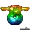
Resolution: 3.62 Å / Resolution method: FSC 0.143 CUT-OFF / Num. of particles: 238017 / Algorithm: FOURIER SPACE / Num. of class averages: 1 / Symmetry type: POINT
Refine LS restraints
Refine-ID
Type
Dev ideal
Number
ELECTRONMICROSCOPY
f_bond_d
0.005
18888
ELECTRONMICROSCOPY
f_angle_d
0.692
25428
ELECTRONMICROSCOPY
f_dihedral_angle_d
24.315
7212
ELECTRONMICROSCOPY
f_chiral_restr
0.045
2832
ELECTRONMICROSCOPY
f_plane_restr
0.005
3252
+
About Yorodumi
-
News
-
Feb 9, 2022. New format data for meta-information of EMDB entries
New format data for meta-information of EMDB entries
Version 3 of the EMDB header file is now the official format.
The previous official version 1.9 will be removed from the archive.
In the structure databanks used in Yorodumi, some data are registered as the other names, "COVID-19 virus" and "2019-nCoV". Here are the details of the virus and the list of structure data.
Jan 31, 2019. EMDB accession codes are about to change! (news from PDBe EMDB page)
EMDB accession codes are about to change! (news from PDBe EMDB page)
The allocation of 4 digits for EMDB accession codes will soon come to an end. Whilst these codes will remain in use, new EMDB accession codes will include an additional digit and will expand incrementally as the available range of codes is exhausted. The current 4-digit format prefixed with “EMD-” (i.e. EMD-XXXX) will advance to a 5-digit format (i.e. EMD-XXXXX), and so on. It is currently estimated that the 4-digit codes will be depleted around Spring 2019, at which point the 5-digit format will come into force.
The EM Navigator/Yorodumi systems omit the EMD- prefix.
Related info.:Q: What is EMD? / ID/Accession-code notation in Yorodumi/EM Navigator
Yorodumi is a browser for structure data from EMDB, PDB, SASBDB, etc.
This page is also the successor to EM Navigator detail page, and also detail information page/front-end page for Omokage search.
The word "yorodu" (or yorozu) is an old Japanese word meaning "ten thousand". "mi" (miru) is to see.
Related info.:EMDB / PDB / SASBDB / Comparison of 3 databanks / Yorodumi Search / Aug 31, 2016. New EM Navigator & Yorodumi / Yorodumi Papers / Jmol/JSmol / Function and homology information / Changes in new EM Navigator and Yorodumi
 Movie
Movie Controller

 Yorodumi
 Open data
 Basic information
 Components
 Keywords
 Function and homology information
 Aquifex aeolicus (bacteria)
 Authors
 Japan, 3items
  Citation
 Structure visualization
 Downloads & links
 Other downloads
 PDBj
PDBj


 Assembly
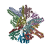
 Sample preparation
 Electron microscopy imaging
 FIELD EMISSION GUN / Accelerating voltage: 200 kV / Illumination mode: FLOOD BEAM
 Processing