 Movie
Movie Controller

[English] 日本語
 Yorodumi
- PDB-7d9t: Structure of human soluble guanylate cyclase in the cinciguat-bou... -
+ Open data

- Basic information
| Entry | Database: PDB / ID: 7d9t | |||||||||||||||
|---|---|---|---|---|---|---|---|---|---|---|---|---|---|---|---|---|
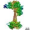
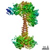
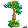
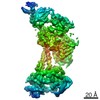
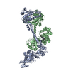
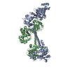
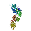
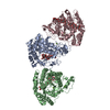
| Title | Structure of human soluble guanylate cyclase in the cinciguat-bound inactive state | |||||||||||||||
 Components Components |
| |||||||||||||||
 Keywords Keywords | SIGNALING PROTEIN / soluble guanylate cyclase | |||||||||||||||
| Function / homology |  Function and homology information Function and homology informationretrograde trans-synaptic signaling by nitric oxide, modulating synaptic transmission / cytidylate cyclase activity / guanylate cyclase complex, soluble / guanylate cyclase / cGMP biosynthetic process / guanylate cyclase activity / presynaptic active zone cytoplasmic component / response to oxygen levels / nitric oxide binding / relaxation of vascular associated smooth muscle ...retrograde trans-synaptic signaling by nitric oxide, modulating synaptic transmission / cytidylate cyclase activity / guanylate cyclase complex, soluble / guanylate cyclase / cGMP biosynthetic process / guanylate cyclase activity / presynaptic active zone cytoplasmic component / response to oxygen levels / nitric oxide binding / relaxation of vascular associated smooth muscle / Nitric oxide stimulates guanylate cyclase / adenylate cyclase activity / : / : / blood circulation / : / Smooth Muscle Contraction / nitric oxide-cGMP-mediated signaling / cellular response to nitric oxide / Hsp90 protein binding / regulation of blood pressure / GABA-ergic synapse / signaling receptor activity / heme binding / GTP binding / protein-containing complex binding / glutamatergic synapse / metal ion binding / cytosol Similarity search - Function | |||||||||||||||
| Biological species |  Homo sapiens (human) Homo sapiens (human) | |||||||||||||||
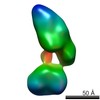
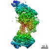
| Method | ELECTRON MICROSCOPY / single particle reconstruction / cryo EM / Resolution: 4.1 Å | |||||||||||||||
 Authors Authors | Chen, L. / Liu, R. / Kang, Y. | |||||||||||||||
| Funding support |  China, 4items China, 4items
| |||||||||||||||
 Citation Citation | Journal: Nat Commun / Year: 2021 Title: Activation mechanism of human soluble guanylate cyclase by stimulators and activators. Authors: Rui Liu / Yunlu Kang / Lei Chen / Abstract: Soluble guanylate cyclase (sGC) is the receptor for nitric oxide (NO) in human. It is an important validated drug target for cardiovascular diseases. sGC can be pharmacologically activated by ...Soluble guanylate cyclase (sGC) is the receptor for nitric oxide (NO) in human. It is an important validated drug target for cardiovascular diseases. sGC can be pharmacologically activated by stimulators and activators. However, the detailed structural mechanisms, through which sGC is recognized and positively modulated by these drugs at high spacial resolution, are poorly understood. Here, we present cryo-electron microscopy structures of human sGC in complex with NO and sGC stimulators, YC-1 and riociguat, and also in complex with the activator cinaciguat. These structures uncover the molecular details of how stimulators interact with residues from both β H-NOX and CC domains, to stabilize sGC in the extended active conformation. In contrast, cinaciguat occupies the haem pocket in the β H-NOX domain and sGC shows both inactive and active conformations. These structures suggest a converged mechanism of sGC activation by pharmacological compounds. | |||||||||||||||
| History |
|
- Structure visualization
| Movie |
Movie viewer |
|---|---|
| Structure viewer | Molecule: MolmilJmol/JSmol |
- Downloads & links
-Download
| PDBx/mmCIF format | 7d9t.cif.gz | 191.7 KB | Display | PDBx/mmCIF format |
|---|---|---|---|---|
| PDB format | pdb7d9t.ent.gz | 140.9 KB | Display | PDB format |
| PDBx/mmJSON format | 7d9t.json.gz | Tree view | PDBx/mmJSON format | |
| Others |  Other downloads Other downloads |
-Validation report
| Arichive directory | https://data.pdbj.org/pub/pdb/validation_reports/d9/7d9tftp://data.pdbj.org/pub/pdb/validation_reports/d9/7d9t | HTTPS FTP |
|---|
-Related structure data
| Related structure data | 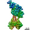 30620MC  7d9rC  7d9sC  7d9uC M: map data used to model this data C: citing same article ( |
|---|---|
| Similar structure data |
-Links
 PDBj
PDBj


- Assembly
| Deposited unit | 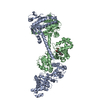
|
|---|---|
| 1 |
|
-Components
| #1: Protein | Mass: 77566.484 Da / Num. of mol.: 1 Source method: isolated from a genetically manipulated source Source: (gene. exp.) Homo sapiens (human) / Gene: GUCY1A1, GUC1A3, GUCSA3, GUCY1A3 / Production host:   Spodoptera frugiperda (fall armyworm) / References: UniProt: Q02108, guanylate cyclase Spodoptera frugiperda (fall armyworm) / References: UniProt: Q02108, guanylate cyclase |
|---|---|
| #2: Protein | Mass: 70599.320 Da / Num. of mol.: 1 Source method: isolated from a genetically manipulated source Source: (gene. exp.) Homo sapiens (human) / Gene: GUCY1B1, GUC1B3, GUCSB3, GUCY1B3 / Production host: Spodoptera frugiperda (fall armyworm) / References: UniProt: Q02153, guanylate cyclase |
| #3: Chemical | ChemComp-Z90 / 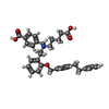  Mass: 565.699 Da / Num. of mol.: 1 / Source method: obtained synthetically / Formula: C36H39NO5 / Feature type: SUBJECT OF INVESTIGATION Mass: 565.699 Da / Num. of mol.: 1 / Source method: obtained synthetically / Formula: C36H39NO5 / Feature type: SUBJECT OF INVESTIGATION |
| Has ligand of interest | Y |
-Experimental details
-Experiment
| Experiment | Method: ELECTRON MICROSCOPY |
|---|---|
| EM experiment | Aggregation state: PARTICLE / 3D reconstruction method: single particle reconstruction |
- Sample preparation
| Component | Name: human soluble guanylate cyclase / Type: COMPLEX / Entity ID: #1-#2 / Source: RECOMBINANT |
|---|---|
| Source (natural) | Organism: Homo sapiens (human) |
| Source (recombinant) | Organism: Spodoptera frugiperda (fall armyworm) |
| Buffer solution | pH: 7.5 |
| Specimen | Embedding applied: NO / Shadowing applied: NO / Staining applied: NO / Vitrification applied: YES |
| Vitrification | Cryogen name: ETHANE |
- Electron microscopy imaging
| Experimental equipment |  Model: Titan Krios / Image courtesy: FEI Company |
|---|---|
| Microscopy | Model: FEI TITAN KRIOS |
| Electron gun | Electron source:  FIELD EMISSION GUN / Accelerating voltage: 300 kV / Illumination mode: FLOOD BEAM FIELD EMISSION GUN / Accelerating voltage: 300 kV / Illumination mode: FLOOD BEAM |
| Electron lens | Mode: BRIGHT FIELD |
| Image recording | Electron dose: 50 e/Å2 / Film or detector model: GATAN K2 QUANTUM (4k x 4k) |
- Processing
| EM software | Name: RELION / Version: 3 / Category: 3D reconstruction |
|---|---|
| CTF correction | Type: NONE |
| 3D reconstruction | Resolution: 4.1 Å / Resolution method: FSC 0.143 CUT-OFF / Num. of particles: 244400 / Symmetry type: POINT |
