 ムービー
ムービー コントローラー


+ データを開く

- 基本情報
基本情報
| 登録情報 | データベース: PDB / ID: 7d7f | |||||||||||||||||||||
|---|---|---|---|---|---|---|---|---|---|---|---|---|---|---|---|---|---|---|---|---|---|---|
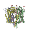
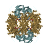
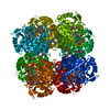
| タイトル | Structure of PKD1L3-CTD/PKD2L1 in calcium-bound state | |||||||||||||||||||||
 要素 要素 |
| |||||||||||||||||||||
 キーワード キーワード | TRANSPORT PROTEIN / Heterotetrameric TRP channel / Calcium / Primary cilia / PKD | |||||||||||||||||||||
| 機能・相同性 |  機能・相同性情報 機能・相同性情報detection of chemical stimulus involved in sensory perception of sour taste / detection of chemical stimulus involved in sensory perception of taste / sensory perception of sour taste / response to water / osmolarity-sensing monoatomic cation channel activity / pH-gated monoatomic ion channel activity / calcium-activated potassium channel activity / cellular response to pH / muscle alpha-actinin binding / detection of mechanical stimulus ...detection of chemical stimulus involved in sensory perception of sour taste / detection of chemical stimulus involved in sensory perception of taste / sensory perception of sour taste / response to water / osmolarity-sensing monoatomic cation channel activity / pH-gated monoatomic ion channel activity / calcium-activated potassium channel activity / cellular response to pH / muscle alpha-actinin binding / detection of mechanical stimulus / cation channel complex / non-motile cilium / calcium-activated cation channel activity / : / smoothened signaling pathway / ciliary membrane / sodium channel activity / alpha-actinin binding / monoatomic cation transmembrane transport / monoatomic cation transport / monoatomic cation channel activity / cellular response to acidic pH / cytoskeletal protein binding / calcium channel complex / potassium ion transmembrane transport / sodium ion transmembrane transport / protein tetramerization / calcium ion transmembrane transport / calcium channel activity / actin cytoskeleton / carbohydrate binding / cytoplasmic vesicle / protein homotetramerization / transmembrane transporter binding / signaling receptor complex / calcium ion binding / endoplasmic reticulum / membrane / identical protein binding / plasma membrane / cytosol 類似検索 - 分子機能 | |||||||||||||||||||||
| 生物種 |  | |||||||||||||||||||||
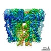
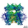
| 手法 | 電子顕微鏡法 / 単粒子再構成法 / クライオ電子顕微鏡法 / 解像度: 3.1 Å | |||||||||||||||||||||
 データ登録者 データ登録者 | Su, Q. / Shi, Y.G. | |||||||||||||||||||||
| 資金援助 |  中国, 2件 中国, 2件
| |||||||||||||||||||||
 引用 引用 | ジャーナル: Nat Commun / 年: 2021 タイトル: Structural basis for Ca activation of the heteromeric PKD1L3/PKD2L1 channel. 著者: Qiang Su / Mengying Chen / Yan Wang / Bin Li / Dan Jing / Xiechao Zhan / Yong Yu / Yigong Shi /  要旨: The heteromeric complex between PKD1L3, a member of the polycystic kidney disease (PKD) protein family, and PKD2L1, also known as TRPP2 or TRPP3, has been a prototype for mechanistic characterization ...The heteromeric complex between PKD1L3, a member of the polycystic kidney disease (PKD) protein family, and PKD2L1, also known as TRPP2 or TRPP3, has been a prototype for mechanistic characterization of heterotetrametric TRP-like channels. Here we show that a truncated PKD1L3/PKD2L1 complex with the C-terminal TRP-fold fragment of PKD1L3 retains both Ca and acid-induced channel activities. Cryo-EM structures of this core heterocomplex with or without supplemented Ca were determined at resolutions of 3.1 Å and 3.4 Å, respectively. The heterotetramer, with a pseudo-symmetric TRP architecture of 1:3 stoichiometry, has an asymmetric selectivity filter (SF) guarded by Lys2069 from PKD1L3 and Asp523 from the three PKD2L1 subunits. Ca-entrance to the SF vestibule is accompanied by a swing motion of Lys2069 on PKD1L3. The S6 of PKD1L3 is pushed inward by the S4-S5 linker of the nearby PKD2L1 (PKD2L1-III), resulting in an elongated intracellular gate which seals the pore domain. Comparison of the apo and Ca-loaded complexes unveils an unprecedented Ca binding site in the extracellular cleft of the voltage-sensing domain (VSD) of PKD2L1-III, but not the other three VSDs. Structure-guided mutagenic studies support this unconventional site to be responsible for Ca-induced channel activation through an allosteric mechanism. | |||||||||||||||||||||
| 履歴 |
|
- 構造の表示
構造の表示
| ムービー |
ムービービューア |
|---|---|
| 構造ビューア | 分子: MolmilJmol/JSmol |
- ダウンロードとリンク
-ダウンロード
| PDBx/mmCIF形式 | 7d7f.cif.gz | 354.1 KB | 表示 | PDBx/mmCIF形式 |
|---|---|---|---|---|
| PDB形式 | pdb7d7f.ent.gz | 282.7 KB | 表示 | PDB形式 |
| PDBx/mmJSON形式 | 7d7f.json.gz | ツリー表示 | PDBx/mmJSON形式 | |
| その他 |  その他のダウンロード その他のダウンロード |
-検証レポート
| アーカイブディレクトリ | https://data.pdbj.org/pub/pdb/validation_reports/d7/7d7fftp://data.pdbj.org/pub/pdb/validation_reports/d7/7d7f | HTTPS FTP |
|---|
-関連構造データ


-リンク
 PDBj
PDBj


- 集合体
集合体
| 登録構造単位 | 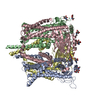
|
|---|---|
| 1 |
|
-要素
| #1: タンパク質 | 分子量: 69234.734 Da / 分子数: 3 / 由来タイプ: 組換発現 / 由来: (組換発現)  Homo sapiens (ヒト) / 参照: UniProt: A2A259 Homo sapiens (ヒト) / 参照: UniProt: A2A259#2: タンパク質 | | 分子量: 62541.914 Da / 分子数: 1 / 由来タイプ: 組換発現 / 由来: (組換発現) Homo sapiens (ヒト) / 参照: UniProt: Q2EG98#3: 糖 | ChemComp-NAG /   タイプ: D-saccharide, beta linking / 分子量: 221.208 Da / 分子数: 10 / 由来タイプ: 合成 / 式: C8H15NO6 タイプ: D-saccharide, beta linking / 分子量: 221.208 Da / 分子数: 10 / 由来タイプ: 合成 / 式: C8H15NO6#4: 化合物 | ChemComp-CA /   分子量: 40.078 Da / 分子数: 5 / 由来タイプ: 合成 / 式: Ca 分子量: 40.078 Da / 分子数: 5 / 由来タイプ: 合成 / 式: Ca研究の焦点であるリガンドがあるか | N | Has protein modification | Y | |
|---|
-実験情報
-実験
| 実験 | 手法: 電子顕微鏡法 |
|---|---|
| EM実験 | 試料の集合状態: PARTICLE / 3次元再構成法: 単粒子再構成法 |
- 試料調製
試料調製
| 構成要素 | 名称: Mouse PKD1L3 in complex with PKD2L1 in presence of 20 mM calcium タイプ: COMPLEX / Entity ID: #1-#2 / 由来: RECOMBINANT |
|---|---|
| 由来(天然) | 生物種: |
| 由来(組換発現) | 生物種: Homo sapiens (ヒト) |
| 緩衝液 | pH: 7.5 |
| 試料 | 濃度: 10 mg/ml / 包埋: NO / シャドウイング: NO / 染色: NO / 凍結: YES |
| 急速凍結 | 凍結剤: ETHANE |
- 電子顕微鏡撮影
| 実験機器 |  モデル: Titan Krios / 画像提供: FEI Company |
|---|---|
| 顕微鏡 | モデル: FEI TITAN KRIOS |
| 電子銃 | 電子線源:  FIELD EMISSION GUN / 加速電圧: 300 kV / 照射モード: FLOOD BEAM FIELD EMISSION GUN / 加速電圧: 300 kV / 照射モード: FLOOD BEAM |
| 電子レンズ | モード: BRIGHT FIELD |
| 撮影 | 電子線照射量: 50 e/Å2 フィルム・検出器のモデル: GATAN K3 BIOQUANTUM (6k x 4k) |
- 解析
解析
| ソフトウェア | 名称: PHENIX / バージョン: 1.17.1_3660: / 分類: 精密化 | ||||||||||||||||||||||||
|---|---|---|---|---|---|---|---|---|---|---|---|---|---|---|---|---|---|---|---|---|---|---|---|---|---|
| EMソフトウェア | 名称: PHENIX / カテゴリ: モデル精密化 | ||||||||||||||||||||||||
| CTF補正 | タイプ: NONE | ||||||||||||||||||||||||
| 3次元再構成 | 解像度: 3.1 Å / 解像度の算出法: FSC 0.143 CUT-OFF / 粒子像の数: 149903 / 対称性のタイプ: POINT | ||||||||||||||||||||||||
| 精密化 | 最高解像度: 3.1 Å | ||||||||||||||||||||||||
| 拘束条件 |
|
