 Movie
Movie Controller


+ Open data

- Basic information
| Entry | Database: PDB / ID: 7cr4 | |||||||||||||||||||||||||||||||||
|---|---|---|---|---|---|---|---|---|---|---|---|---|---|---|---|---|---|---|---|---|---|---|---|---|---|---|---|---|---|---|---|---|---|---|
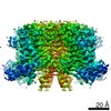
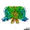
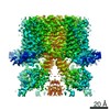
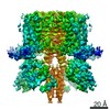
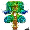
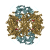
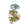
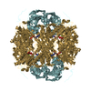
| Title | human KCNQ2-CaM in complex with ztz240 | |||||||||||||||||||||||||||||||||
 Components Components |
| |||||||||||||||||||||||||||||||||
 Keywords Keywords | TRANSPORT PROTEIN / ion channel | |||||||||||||||||||||||||||||||||
| Function / homology |  Function and homology information Function and homology informationCASP4 inflammasome assembly / transporter inhibitor activity / : / axon initial segment / type 3 metabotropic glutamate receptor binding / Voltage gated Potassium channels / node of Ranvier / Enterobacterial factors antagonize host defense / Interaction between L1 and Ankyrins / ankyrin binding ...CASP4 inflammasome assembly / transporter inhibitor activity / : / axon initial segment / type 3 metabotropic glutamate receptor binding / Voltage gated Potassium channels / node of Ranvier / Enterobacterial factors antagonize host defense / Interaction between L1 and Ankyrins / ankyrin binding / voltage-gated monoatomic cation channel activity / negative regulation of high voltage-gated calcium channel activity / response to corticosterone / regulation of synaptic vesicle exocytosis / negative regulation of calcium ion export across plasma membrane / regulation of cardiac muscle cell action potential / presynaptic endocytosis / calcineurin-mediated signaling / nitric-oxide synthase binding / regulation of cell communication by electrical coupling involved in cardiac conduction / adenylate cyclase binding / protein phosphatase activator activity / action potential / regulation of synaptic vesicle endocytosis / voltage-gated potassium channel activity / detection of calcium ion / postsynaptic cytosol / catalytic complex / regulation of cardiac muscle contraction / phosphatidylinositol 3-kinase binding / presynaptic cytosol / regulation of release of sequestered calcium ion into cytosol by sarcoplasmic reticulum / titin binding / regulation of cardiac muscle contraction by regulation of the release of sequestered calcium ion / voltage-gated potassium channel complex / calcium channel complex / substantia nigra development / potassium ion transmembrane transport / regulation of heart rate / calyx of Held / response to amphetamine / nitric-oxide synthase regulator activity / adenylate cyclase activator activity / protein serine/threonine kinase activator activity / regulation of cytokinesis / spindle microtubule / sarcomere / calcium channel regulator activity / response to calcium ion / Schaffer collateral - CA1 synapse / mitochondrial membrane / long-term synaptic potentiation / spindle pole / calcium-dependent protein binding / myelin sheath / nervous system development / sperm midpiece / synaptic vesicle membrane / growth cone / chemical synaptic transmission / vesicle / transmembrane transporter binding / calmodulin binding / G protein-coupled receptor signaling pathway / protein domain specific binding / calcium ion binding / centrosome / synapse / protein kinase binding / chromatin / protein-containing complex / membrane / nucleus / plasma membrane / cytosol / cytoplasm Similarity search - Function | |||||||||||||||||||||||||||||||||
| Biological species |  Homo sapiens (human) Homo sapiens (human) | |||||||||||||||||||||||||||||||||
| Method | ELECTRON MICROSCOPY / single particle reconstruction / cryo EM / Resolution: 3.9 Å | |||||||||||||||||||||||||||||||||
 Authors Authors | Li, X. / Lv, D. / Wang, J. / Ye, S. / Guo, J. | |||||||||||||||||||||||||||||||||
| Funding support |  China, 2items China, 2items
| |||||||||||||||||||||||||||||||||
 Citation Citation | Journal: Cell Res / Year: 2021 Title: Molecular basis for ligand activation of the human KCNQ2 channel. Authors: Xiaoxiao Li / Qiansen Zhang / Peipei Guo / Jie Fu / Lianghe Mei / Dashuai Lv / Jiangqin Wang / Dongwu Lai / Sheng Ye / Huaiyu Yang / Jiangtao Guo / Abstract: The voltage-gated potassium channel KCNQ2 is responsible for M-current in neurons and is an important drug target to treat epilepsy, pain and several other diseases related to neuronal hyper- ...The voltage-gated potassium channel KCNQ2 is responsible for M-current in neurons and is an important drug target to treat epilepsy, pain and several other diseases related to neuronal hyper-excitability. A list of synthetic compounds have been developed to directly activate KCNQ2, yet our knowledge of their activation mechanism is limited, due to lack of high-resolution structures. Here, we report cryo-electron microscopy (cryo-EM) structures of the human KCNQ2 determined in apo state and in complex with two activators, ztz240 or retigabine, which activate KCNQ2 through different mechanisms. The activator-bound structures, along with electrophysiology analysis, reveal that ztz240 binds at the voltage-sensing domain and directly stabilizes it at the activated state, whereas retigabine binds at the pore domain and activates the channel by an allosteric modulation. By accurately defining ligand-binding sites, these KCNQ2 structures not only reveal different ligand recognition and activation mechanisms, but also provide a structural basis for drug optimization and design. | |||||||||||||||||||||||||||||||||
| History |
|
- Structure visualization
| Movie |
Movie viewer |
|---|---|
| Structure viewer | Molecule: MolmilJmol/JSmol |
- Downloads & links
-Download
| PDBx/mmCIF format | 7cr4.cif.gz | 369 KB | Display | PDBx/mmCIF format |
|---|---|---|---|---|
| PDB format | pdb7cr4.ent.gz | 294.4 KB | Display | PDB format |
| PDBx/mmJSON format | 7cr4.json.gz | Tree view | PDBx/mmJSON format | |
| Others |  Other downloads Other downloads |
-Validation report
| Arichive directory | https://data.pdbj.org/pub/pdb/validation_reports/cr/7cr4ftp://data.pdbj.org/pub/pdb/validation_reports/cr/7cr4 | HTTPS FTP |
|---|
-Related structure data
| Related structure data | 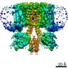 30447MC 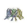 7cr0C 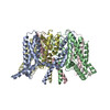 7cr1C 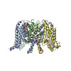 7cr2C 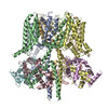 7cr3C 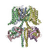 7cr7C C: citing same article ( M: map data used to model this data |
|---|---|
| Similar structure data |


-Links
 PDBj
PDBj
- Assembly
| Deposited unit | 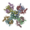
|
|---|---|
| 1 |
|
-Components
| #1: Protein | Mass: 73627.812 Da / Num. of mol.: 4 Source method: isolated from a genetically manipulated source Source: (gene. exp.) Homo sapiens (human) / Gene: KCNQ2 / Cell line (production host): HEK293 / Production host: Homo sapiens (human) / References: UniProt: O43526#2: Protein | Mass: 16852.545 Da / Num. of mol.: 4 / Source method: isolated from a natural source / Source: (natural) Homo sapiens (human) / References: UniProt: P0DP25#3: Chemical | ChemComp-GB9 / 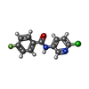  Mass: 250.656 Da / Num. of mol.: 4 / Source method: obtained synthetically / Formula: C12H8ClFN2O / Feature type: SUBJECT OF INVESTIGATION Mass: 250.656 Da / Num. of mol.: 4 / Source method: obtained synthetically / Formula: C12H8ClFN2O / Feature type: SUBJECT OF INVESTIGATIONHas ligand of interest | Y | Has protein modification | N | |
|---|
-Experimental details
-Experiment
| Experiment | Method: ELECTRON MICROSCOPY |
|---|---|
| EM experiment | Aggregation state: PARTICLE / 3D reconstruction method: single particle reconstruction |
- Sample preparation
| Component | Name: voltage-gated potassium channel KCNQ2 / Type: COMPLEX / Entity ID: #1 / Source: RECOMBINANT |
|---|---|
| Source (natural) | Organism: Homo sapiens (human) |
| Source (recombinant) | Organism: Homo sapiens (human) |
| Buffer solution | pH: 8 |
| Specimen | Embedding applied: NO / Shadowing applied: NO / Staining applied: NO / Vitrification applied: YES |
| Vitrification | Cryogen name: ETHANE |
- Electron microscopy imaging
| Experimental equipment |  Model: Titan Krios / Image courtesy: FEI Company |
|---|---|
| Microscopy | Model: FEI TITAN KRIOS |
| Electron gun | Electron source:  FIELD EMISSION GUN / Accelerating voltage: 300 kV / Illumination mode: FLOOD BEAM FIELD EMISSION GUN / Accelerating voltage: 300 kV / Illumination mode: FLOOD BEAM |
| Electron lens | Mode: BRIGHT FIELD |
| Image recording | Electron dose: 1.556 e/Å2 / Film or detector model: GATAN K2 SUMMIT (4k x 4k) |
- Processing
| Software | Name: PHENIX / Version: 1.15.2_3472: / Classification: refinement | ||||||||||||||||||||||||
|---|---|---|---|---|---|---|---|---|---|---|---|---|---|---|---|---|---|---|---|---|---|---|---|---|---|
| EM software | Name: PHENIX / Category: model refinement | ||||||||||||||||||||||||
| CTF correction | Type: PHASE FLIPPING AND AMPLITUDE CORRECTION | ||||||||||||||||||||||||
| 3D reconstruction | Resolution: 3.9 Å / Resolution method: FSC 0.143 CUT-OFF / Num. of particles: 86371 / Symmetry type: POINT | ||||||||||||||||||||||||
| Refine LS restraints |
|
