 Movie
Movie Controller


+ Open data

- Basic information
| Entry | Database: PDB / ID: 7b0u | ||||||
|---|---|---|---|---|---|---|---|
| Title | Stressosome complex from Listeria innocua | ||||||
 Components Components |
| ||||||
 Keywords Keywords | SIGNALING PROTEIN / Stressosome / stress sensing / general stress response / phosphorylation cascade | ||||||
| Function / homology |  Function and homology information Function and homology information | ||||||
| Biological species | Listeria innocua serovar 6a | ||||||
| Method | ELECTRON MICROSCOPY / single particle reconstruction / cryo EM / Resolution: 3.86 Å | ||||||
 Authors Authors | Miksys, A. / Fu, L. / Madej, M.G. / Ziegler, C. | ||||||
| Funding support |  Germany, 1items Germany, 1items
| ||||||
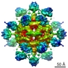
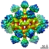
 Citation Citation | Journal: Commun Biol / Year: 2022 Title: Molecular insights into intra-complex signal transmission during stressosome activation. Authors: Algirdas Miksys / Lifei Fu / M Gregor Madej / Duarte N Guerreiro / Susann Kaltwasser / Maria Conway / Sema Ejder / Astrid Bruckmann / Jon Marles-Wright / Richard J Lewis / Conor O'Byrne / ...Authors: Algirdas Miksys / Lifei Fu / M Gregor Madej / Duarte N Guerreiro / Susann Kaltwasser / Maria Conway / Sema Ejder / Astrid Bruckmann / Jon Marles-Wright / Richard J Lewis / Conor O'Byrne / Jan Pané-Farré / Christine Ziegler /   Abstract: The stressosome is a pseudo-icosahedral megadalton bacterial stress-sensing protein complex consisting of several copies of two STAS-domain proteins, RsbR and RsbS, and the kinase RsbT. Upon ...The stressosome is a pseudo-icosahedral megadalton bacterial stress-sensing protein complex consisting of several copies of two STAS-domain proteins, RsbR and RsbS, and the kinase RsbT. Upon perception of environmental stress multiple copies of RsbT are released from the surface of the stressosome. Free RsbT activates downstream proteins to elicit a global cellular response, such as the activation of the general stress response in Gram-positive bacteria. The molecular events triggering RsbT release from the stressosome surface remain poorly understood. Here we present the map of Listeria innocua RsbR1/RsbS complex at resolutions of 3.45 Å for the STAS domain core in icosahedral symmetry and of 3.87 Å for the STAS domain and N-terminal sensors in D2 symmetry, respectively. The structure reveals a conformational change in the STAS domain linked to phosphorylation in RsbR. Docking studies indicate that allosteric RsbT binding to the conformationally flexible N-terminal sensor domain of RsbR affects the affinity of RsbS towards RsbT. Our results bring to focus the molecular events within the stressosome complex and further our understanding of this ubiquitous signaling hub. | ||||||
| History |
|
- Structure visualization
| Movie |
Movie viewer |
|---|---|
| Structure viewer | Molecule: MolmilJmol/JSmol |
- Downloads & links
-Download
| PDBx/mmCIF format | 7b0u.cif.gz | 2.1 MB | Display | PDBx/mmCIF format |
|---|---|---|---|---|
| PDB format | pdb7b0u.ent.gz | Display | PDB format | |
| PDBx/mmJSON format | 7b0u.json.gz | Tree view | PDBx/mmJSON format | |
| Others |  Other downloads Other downloads |
-Validation report
| Arichive directory | https://data.pdbj.org/pub/pdb/validation_reports/b0/7b0uftp://data.pdbj.org/pub/pdb/validation_reports/b0/7b0u | HTTPS FTP |
|---|
-Related structure data
| Related structure data | 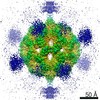 11971MC M: map data used to model this data C: citing same article ( |
|---|---|
| Similar structure data |


-Links
 PDBj
PDBj


- Assembly
| Deposited unit | 
|
|---|---|
| 1 |
|
-Components
| #1: Protein | Mass: 31704.586 Da / Num. of mol.: 16 Source method: isolated from a genetically manipulated source Source: (gene. exp.)  Listeria innocua serovar 6a (strain ATCC BAA-680 / CLIP 11262) (bacteria) Listeria innocua serovar 6a (strain ATCC BAA-680 / CLIP 11262) (bacteria)Strain: ATCC BAA-680 / CLIP 11262 / Gene: RsbR / Production host: #2: Protein | Mass: 31784.566 Da / Num. of mol.: 16 Source method: isolated from a genetically manipulated source Source: (gene. exp.) Listeria innocua serovar 6a (strain ATCC BAA-680 / CLIP 11262) (bacteria)Strain: ATCC BAA-680 / CLIP 11262 / Gene: RsbR / Production host: #3: Protein | Mass: 12606.707 Da / Num. of mol.: 20 Source method: isolated from a genetically manipulated source Source: (gene. exp.) Listeria innocua serovar 6a (strain ATCC BAA-680 / CLIP 11262) (bacteria)Strain: ATCC BAA-680 / CLIP 11262 / Gene: rsbS / Production host: #4: Protein | Mass: 31864.547 Da / Num. of mol.: 4 / Mutation: T175/241-TPO Source method: isolated from a genetically manipulated source Source: (gene. exp.) Listeria innocua serovar 6a (strain ATCC BAA-680 / CLIP 11262) (bacteria)Strain: ATCC BAA-680 / CLIP 11262 / Gene: RsbR / Production host: #5: Protein | Mass: 31784.566 Da / Num. of mol.: 4 / Mutation: T175-TPO Source method: isolated from a genetically manipulated source Source: (gene. exp.) Listeria innocua serovar 6a (strain ATCC BAA-680 / CLIP 11262) (bacteria)Strain: ATCC BAA-680 / CLIP 11262 / Gene: RsbR / Production host: Has ligand of interest | N | Has protein modification | Y | |
|---|
-Experimental details
-Experiment
| Experiment | Method: ELECTRON MICROSCOPY |
|---|---|
| EM experiment | Aggregation state: PARTICLE / 3D reconstruction method: single particle reconstruction |
- Sample preparation
| Component | Name: stressosome complex RsbR and RsbS / Type: COMPLEX / Entity ID: all / Source: RECOMBINANT | |||||||||||||||
|---|---|---|---|---|---|---|---|---|---|---|---|---|---|---|---|---|
| Molecular weight | Value: 1.5 MDa / Experimental value: NO | |||||||||||||||
| Source (natural) | Organism: Listeria innocua Clip11262 (bacteria) | |||||||||||||||
| Source (recombinant) | Organism: | |||||||||||||||
| Buffer solution | pH: 8.5 / Details: pH 8.5 | |||||||||||||||
| Buffer component |
| |||||||||||||||
| Specimen | Conc.: 0.6 mg/ml / Embedding applied: NO / Shadowing applied: NO / Staining applied: NO / Vitrification applied: YES | |||||||||||||||
| Specimen support | Grid material: COPPER / Grid type: Quantifoil | |||||||||||||||
| Vitrification | Instrument: FEI VITROBOT MARK IV / Cryogen name: ETHANE / Humidity: 100 % / Chamber temperature: 277 K |
- Electron microscopy imaging
| Experimental equipment |  Model: Titan Krios / Image courtesy: FEI Company |
|---|---|
| Microscopy | Model: TFS KRIOS |
| Electron gun | Electron source:  FIELD EMISSION GUN / Accelerating voltage: 300 kV / Illumination mode: FLOOD BEAM FIELD EMISSION GUN / Accelerating voltage: 300 kV / Illumination mode: FLOOD BEAM |
| Electron lens | Mode: BRIGHT FIELD / Cs: 2.7 mm |
| Specimen holder | Cryogen: NITROGEN / Specimen holder model: FEI TITAN KRIOS AUTOGRID HOLDER |
| Image recording | Electron dose: 50 e/Å2 / Detector mode: INTEGRATING / Film or detector model: FEI FALCON III (4k x 4k) |
- Processing
| Software | Name: PHENIX / Version: 1.19rc5_4047: phenix.real_space_refine / Classification: refinement | ||||||||||||||||
|---|---|---|---|---|---|---|---|---|---|---|---|---|---|---|---|---|---|
| EM software |
| ||||||||||||||||
| CTF correction | Type: PHASE FLIPPING AND AMPLITUDE CORRECTION | ||||||||||||||||
| Symmetry | Point symmetry: D2 (2x2 fold dihedral) | ||||||||||||||||
| 3D reconstruction | Resolution: 3.86 Å / Resolution method: FSC 0.143 CUT-OFF / Num. of particles: 32031 / Symmetry type: POINT | ||||||||||||||||
| Atomic model building | Protocol: AB INITIO MODEL / Space: REAL / Target criteria: Correlation coefficient |
