 Movie
Movie Controller


+ Open data

- Basic information
| Entry | Database: PDB / ID: 7wzx | ||||||
|---|---|---|---|---|---|---|---|
| Title | The structure of a Twitch Radical SAM Dehydrogenase SpeY | ||||||
 Components Components | 4Fe-4S cluster-binding domain-containing protein | ||||||
 Keywords Keywords | BIOSYNTHETIC PROTEIN / Spectinomycin / Twitch Radical SAM Dehydrogenase / Epimerase | ||||||
| Function / homology |  Function and homology information Function and homology informationcatalytic activity / 4 iron, 4 sulfur cluster binding / metal ion binding Similarity search - Function | ||||||
| Biological species |  Streptomyces spectabilis (bacteria) Streptomyces spectabilis (bacteria) | ||||||
| Method |  X-RAY DIFFRACTION / SYNCHROTRON / MOLECULAR REPLACEMENT / Resolution: 1.98001304567 Å X-RAY DIFFRACTION / SYNCHROTRON / MOLECULAR REPLACEMENT / Resolution: 1.98001304567 Å | ||||||
 Authors Authors | Hou, X.L. / Zhou, J.H. | ||||||
| Funding support | 1items
| ||||||
 Citation Citation | Journal: J.Am.Chem.Soc. / Year: 2022 Title: Dioxane Bridge Formation during the Biosynthesis of Spectinomycin Involves a Twitch Radical S -Adenosyl Methionine Dehydrogenase That May Have Evolved from an Epimerase. Authors: Zhang, J. / Hou, X. / Chen, Z. / Ko, Y. / Ruszczycky, M.W. / Chen, Y. / Zhou, J. / Liu, H.W. | ||||||
| History |
|
- Structure visualization
| Structure viewer | Molecule: MolmilJmol/JSmol |
|---|
- Downloads & links
-Download
| PDBx/mmCIF format | 7wzx.cif.gz | 173.3 KB | Display | PDBx/mmCIF format |
|---|---|---|---|---|
| PDB format | pdb7wzx.ent.gz | 112.7 KB | Display | PDB format |
| PDBx/mmJSON format | 7wzx.json.gz | Tree view | PDBx/mmJSON format | |
| Others |  Other downloads Other downloads |
-Validation report
| Arichive directory | https://data.pdbj.org/pub/pdb/validation_reports/wz/7wzxftp://data.pdbj.org/pub/pdb/validation_reports/wz/7wzx | HTTPS FTP |
|---|
-Related structure data
| Related structure data | 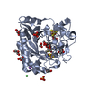 7wzvSC 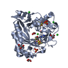 7x0bC S: Starting model for refinement C: citing same article ( |
|---|---|
| Similar structure data |
-Links
 PDBj
PDBj
- Assembly
| Deposited unit | 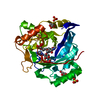
| ||||||||||||
|---|---|---|---|---|---|---|---|---|---|---|---|---|---|
| 1 |
| ||||||||||||
| Unit cell |
| ||||||||||||
| Components on special symmetry positions |
|
-Components
-Protein , 1 types, 1 molecules A
| #1: Protein | Mass: 36014.637 Da / Num. of mol.: 1 Source method: isolated from a genetically manipulated source Source: (gene. exp.) Streptomyces spectabilis (bacteria) / Gene: speY, FH965_34950 / Production host: |
|---|
-Non-polymers , 6 types, 91 molecules 


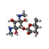


| #2: Chemical |  Mass: 351.640 Da / Num. of mol.: 2 / Source method: obtained synthetically / Formula: Fe4S4 Mass: 351.640 Da / Num. of mol.: 2 / Source method: obtained synthetically / Formula: Fe4S4#3: Chemical | ChemComp-SAM / |  Mass: 398.437 Da / Num. of mol.: 1 / Source method: obtained synthetically / Formula: C15H22N6O5S Mass: 398.437 Da / Num. of mol.: 1 / Source method: obtained synthetically / Formula: C15H22N6O5S#4: Chemical | ChemComp-GOL /  Mass: 92.094 Da / Num. of mol.: 4 / Source method: obtained synthetically / Formula: C3H8O3 Mass: 92.094 Da / Num. of mol.: 4 / Source method: obtained synthetically / Formula: C3H8O3#5: Chemical | ChemComp-7PK / ( |  Mass: 334.365 Da / Num. of mol.: 1 / Source method: obtained synthetically / Formula: C14H26N2O7 / Feature type: SUBJECT OF INVESTIGATION Mass: 334.365 Da / Num. of mol.: 1 / Source method: obtained synthetically / Formula: C14H26N2O7 / Feature type: SUBJECT OF INVESTIGATION#6: Chemical |  Mass: 96.063 Da / Num. of mol.: 2 / Source method: obtained synthetically / Formula: SO4 Mass: 96.063 Da / Num. of mol.: 2 / Source method: obtained synthetically / Formula: SO4#7: Water | ChemComp-HOH / | Mass: 18.015 Da / Num. of mol.: 81 / Source method: isolated from a natural source / Formula: H2O |
|---|
-Details
| Has ligand of interest | Y |
|---|
-Experimental details
-Experiment
| Experiment | Method: X-RAY DIFFRACTION / Number of used crystals: 1 |
|---|
- Sample preparation
| Crystal | Density Matthews: 2.54 Å3/Da / Density % sol: 51.49 % |
|---|---|
| Crystal grow | Temperature: 293 K / Method: vapor diffusion, sitting drop Details: 0.5 M Sodium cacodylate trihydrate (pH 6.0), 1.8 M Ammonium sulfate and 0.05 M Magnesium acetate tetrahydrate |
-Data collection
| Diffraction | Mean temperature: 100 K / Serial crystal experiment: N |
|---|---|
| Diffraction source | Source: SYNCHROTRON / Site: SSRF  / Beamline: BL19U1 / Wavelength: 0.97846 Å / Beamline: BL19U1 / Wavelength: 0.97846 Å |
| Detector | Type: DECTRIS PILATUS 6M / Detector: PIXEL / Date: Jan 30, 2021 |
| Radiation | Protocol: SINGLE WAVELENGTH / Monochromatic (M) / Laue (L): M / Scattering type: x-ray |
| Radiation wavelength | Wavelength: 0.97846 Å / Relative weight: 1 |
| Reflection | Resolution: 1.98→44.63 Å / Num. obs: 26783 / % possible obs: 100 % / Redundancy: 37.9 % / Biso Wilson estimate: 34.0387505161 Å2 / CC1/2: 1 / Net I/σ(I): 42.3 |
| Reflection shell | Resolution: 1.98→2.03 Å / Rmerge(I) obs: 0.963 / Num. unique obs: 1844 / CC1/2: 0.949 |
- Processing
| Software |
| ||||||||||||||||||||||||||||||||||||||||||||||||||||||||||||||||||||||
|---|---|---|---|---|---|---|---|---|---|---|---|---|---|---|---|---|---|---|---|---|---|---|---|---|---|---|---|---|---|---|---|---|---|---|---|---|---|---|---|---|---|---|---|---|---|---|---|---|---|---|---|---|---|---|---|---|---|---|---|---|---|---|---|---|---|---|---|---|---|---|---|
| Refinement | Method to determine structure: MOLECULAR REPLACEMENT Starting model: 7WZV Resolution: 1.98001304567→41.2158933488 Å / SU ML: 0.250812795788 / Cross valid method: FREE R-VALUE / σ(F): 1.35002947799 / Phase error: 24.4586368563 Stereochemistry target values: GeoStd + Monomer Library + CDL v1.2
| ||||||||||||||||||||||||||||||||||||||||||||||||||||||||||||||||||||||
| Solvent computation | Shrinkage radii: 0.9 Å / VDW probe radii: 1.11 Å / Solvent model: FLAT BULK SOLVENT MODEL | ||||||||||||||||||||||||||||||||||||||||||||||||||||||||||||||||||||||
| Displacement parameters | Biso mean: 49.3943035328 Å2 | ||||||||||||||||||||||||||||||||||||||||||||||||||||||||||||||||||||||
| Refinement step | Cycle: LAST / Resolution: 1.98001304567→41.2158933488 Å
| ||||||||||||||||||||||||||||||||||||||||||||||||||||||||||||||||||||||
| Refine LS restraints |
| ||||||||||||||||||||||||||||||||||||||||||||||||||||||||||||||||||||||
| LS refinement shell |
| ||||||||||||||||||||||||||||||||||||||||||||||||||||||||||||||||||||||
| Refinement TLS params. | Method: refined / Origin x: 2.63957130723 Å / Origin y: 36.4786141394 Å / Origin z: 2.53794544531 Å
| ||||||||||||||||||||||||||||||||||||||||||||||||||||||||||||||||||||||
| Refinement TLS group | Selection details: all |
