+ Open data
- Basic information
| Entry | Database: PDB / ID: 7wmv | ||||||||||||
|---|---|---|---|---|---|---|---|---|---|---|---|---|---|
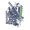
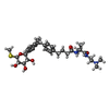
| Title | Structure of human SGLT1-MAP17 complex bound with LX2761 | ||||||||||||
 Components Components |
| ||||||||||||
 Keywords Keywords | PROTEIN TRANSPORT / glucose transporter / SGLT / sodium glucose transporter / membrane protein | ||||||||||||
| Function / homology |  Function and homology information Function and homology informationmyo-inositol:sodium symporter activity / pentose transmembrane transporter activity / galactose:sodium symporter activity / pentose transmembrane transport / myo-inositol transport / intestinal hexose absorption / Defective SLC5A1 causes congenital glucose/galactose malabsorption (GGM) / Intestinal hexose absorption / fucose transmembrane transport / fucose transmembrane transporter activity ...myo-inositol:sodium symporter activity / pentose transmembrane transporter activity / galactose:sodium symporter activity / pentose transmembrane transport / myo-inositol transport / intestinal hexose absorption / Defective SLC5A1 causes congenital glucose/galactose malabsorption (GGM) / Intestinal hexose absorption / fucose transmembrane transport / fucose transmembrane transporter activity / intestinal D-glucose absorption / galactose transmembrane transporter activity / galactose transmembrane transport / alpha-glucoside transport / alpha-glucoside transmembrane transporter activity / D-glucose:sodium symporter activity / water transmembrane transporter activity / renal D-glucose absorption / D-glucose import across plasma membrane / Cellular hexose transport / D-glucose transmembrane transporter activity / D-glucose transmembrane transport / transepithelial water transport / sodium ion import across plasma membrane / sodium ion transport / intracellular vesicle / transport across blood-brain barrier / brush border membrane / nuclear membrane / early endosome / apical plasma membrane / perinuclear region of cytoplasm / Golgi apparatus / extracellular exosome / plasma membrane Similarity search - Function | ||||||||||||
| Biological species |  Homo sapiens (human) Homo sapiens (human) | ||||||||||||
| Method | ELECTRON MICROSCOPY / single particle reconstruction / cryo EM / Resolution: 3.2 Å | ||||||||||||
 Authors Authors | Chen, L. / Niu, Y. / Cui, W. | ||||||||||||
| Funding support |  China, 3items China, 3items
| ||||||||||||
 Citation Citation |  Journal: Nat Commun / Year: 2022 Journal: Nat Commun / Year: 2022Title: Structural mechanism of SGLT1 inhibitors. Authors: Yange Niu / Wenhao Cui / Rui Liu / Sanshan Wang / Han Ke / Xiaoguang Lei / Lei Chen /  Abstract: Sodium glucose co-transporters (SGLT) harness the electrochemical gradient of sodium to drive the uphill transport of glucose across the plasma membrane. Human SGLT1 (hSGLT1) plays a key role in ...Sodium glucose co-transporters (SGLT) harness the electrochemical gradient of sodium to drive the uphill transport of glucose across the plasma membrane. Human SGLT1 (hSGLT1) plays a key role in sugar uptake from food and its inhibitors show promise in the treatment of several diseases. However, the inhibition mechanism for hSGLT1 remains elusive. Here, we present the cryo-EM structure of the hSGLT1-MAP17 hetero-dimeric complex in the presence of the high-affinity inhibitor LX2761. LX2761 locks the transporter in an outward-open conformation by wedging inside the substrate-binding site and the extracellular vestibule of hSGLT1. LX2761 blocks the putative water permeation pathway of hSGLT1. The structure also uncovers the conformational changes of hSGLT1 during transitions from outward-open to inward-open states. | ||||||||||||
| History |
|
- Structure visualization
| Structure viewer | Molecule:  Molmil Molmil Jmol/JSmol Jmol/JSmol |
|---|
- Downloads & links
- Download
| PDBx/mmCIF format |  7wmv.cif.gz 7wmv.cif.gz | 135.8 KB | Display |  PDBx/mmCIF format PDBx/mmCIF format |
|---|---|---|---|---|
| PDB format |  pdb7wmv.ent.gz pdb7wmv.ent.gz | 102.5 KB | Display |  PDB format PDB format |
| PDBx/mmJSON format |  7wmv.json.gz 7wmv.json.gz | Tree view |  PDBx/mmJSON format PDBx/mmJSON format | |
| Others |  Other downloads Other downloads |
-Validation report
| Arichive directory |  https://data.pdbj.org/pub/pdb/validation_reports/wm/7wmv https://data.pdbj.org/pub/pdb/validation_reports/wm/7wmv ftp://data.pdbj.org/pub/pdb/validation_reports/wm/7wmv ftp://data.pdbj.org/pub/pdb/validation_reports/wm/7wmv | HTTPS FTP |
|---|
-Related structure data
| Related structure data |  32617MC M: map data used to model this data C: citing same article ( |
|---|---|
| Similar structure data | Similarity search - Function & homology  F&H Search F&H Search |
- Links
Links
- Assembly
| Deposited unit | 
|
|---|---|
| 1 |
|
- Components
| #1: Protein | Mass: 73557.703 Da / Num. of mol.: 1 Source method: isolated from a genetically manipulated source Source: (gene. exp.)  Homo sapiens (human) / Gene: SLC5A1, NAGT, SGLT1 / Production host: Homo sapiens (human) / Gene: SLC5A1, NAGT, SGLT1 / Production host:  Homo sapiens (human) / References: UniProt: P13866 Homo sapiens (human) / References: UniProt: P13866 |
|---|---|
| #2: Protein | Mass: 12265.092 Da / Num. of mol.: 1 Source method: isolated from a genetically manipulated source Source: (gene. exp.)  Homo sapiens (human) / Gene: PDZK1IP1, MAP17 / Production host: Homo sapiens (human) / Gene: PDZK1IP1, MAP17 / Production host:  Homo sapiens (human) / References: UniProt: Q13113 Homo sapiens (human) / References: UniProt: Q13113 |
| #3: Chemical | ChemComp-1YI / |
| Has ligand of interest | Y |
| Has protein modification | Y |
-Experimental details
-Experiment
| Experiment | Method: ELECTRON MICROSCOPY |
|---|---|
| EM experiment | Aggregation state: PARTICLE / 3D reconstruction method: single particle reconstruction |
- Sample preparation
| Component | Name: human SGLT1-MAP17 complex / Type: COMPLEX / Entity ID: #1-#2 / Source: RECOMBINANT |
|---|---|
| Source (natural) | Organism:  Homo sapiens (human) Homo sapiens (human) |
| Source (recombinant) | Organism:  Homo sapiens (human) Homo sapiens (human) |
| Buffer solution | pH: 7.5 |
| Specimen | Embedding applied: NO / Shadowing applied: NO / Staining applied: NO / Vitrification applied: YES |
| Vitrification | Cryogen name: ETHANE |
- Electron microscopy imaging
| Experimental equipment |  Model: Titan Krios / Image courtesy: FEI Company |
|---|---|
| Microscopy | Model: FEI TITAN KRIOS |
| Electron gun | Electron source:  FIELD EMISSION GUN / Accelerating voltage: 300 kV / Illumination mode: SPOT SCAN FIELD EMISSION GUN / Accelerating voltage: 300 kV / Illumination mode: SPOT SCAN |
| Electron lens | Mode: BRIGHT FIELD / Nominal defocus max: 1800 nm / Nominal defocus min: 1500 nm |
| Image recording | Electron dose: 50 e/Å2 / Film or detector model: GATAN K2 SUMMIT (4k x 4k) |
- Processing
| EM software | Name: cryoSPARC / Version: v3.1.0 / Category: 3D reconstruction |
|---|---|
| CTF correction | Type: NONE |
| 3D reconstruction | Resolution: 3.2 Å / Resolution method: FSC 0.143 CUT-OFF / Num. of particles: 12133 / Symmetry type: POINT |
 Movie
Movie Controller


 PDBj
PDBj


