 Movie
Movie Controller

[English] 日本語
 Yorodumi
- PDB-7qur: SARS-CoV-2 Spike with ethylbenzamide-tri-iodo Siallyllactose, C3 ... -
+ Open data

- Basic information
| Entry | Database: PDB / ID: 7qur | |||||||||||||||||||||||||||||||||||||||
|---|---|---|---|---|---|---|---|---|---|---|---|---|---|---|---|---|---|---|---|---|---|---|---|---|---|---|---|---|---|---|---|---|---|---|---|---|---|---|---|---|
| Title | SARS-CoV-2 Spike with ethylbenzamide-tri-iodo Siallyllactose, C3 symmetry | |||||||||||||||||||||||||||||||||||||||
 Components Components | Spike glycoprotein,Fibritin | |||||||||||||||||||||||||||||||||||||||
 Keywords Keywords | VIRAL PROTEIN / SARS-CoV-2 Spike | |||||||||||||||||||||||||||||||||||||||
| Function / homology |  Function and homology information Function and homology informationvirion component / symbiont-mediated disruption of host tissue / Maturation of spike protein / Translation of Structural Proteins / Virion Assembly and Release / host cell surface / host extracellular region / symbiont-mediated-mediated suppression of host tetherin activity / Induction of Cell-Cell Fusion / structural constituent of virion ...virion component / symbiont-mediated disruption of host tissue / Maturation of spike protein / Translation of Structural Proteins / Virion Assembly and Release / host cell surface / host extracellular region / symbiont-mediated-mediated suppression of host tetherin activity / Induction of Cell-Cell Fusion / structural constituent of virion / positive regulation of viral entry into host cell / membrane fusion / host cell endoplasmic reticulum-Golgi intermediate compartment membrane / Attachment and Entry / entry receptor-mediated virion attachment to host cell / receptor-mediated virion attachment to host cell / host cell surface receptor binding / symbiont-mediated suppression of host innate immune response / endocytosis involved in viral entry into host cell / receptor ligand activity / fusion of virus membrane with host plasma membrane / fusion of virus membrane with host endosome membrane / viral envelope / symbiont entry into host cell / virion attachment to host cell / host cell plasma membrane / SARS-CoV-2 activates/modulates innate and adaptive immune responses / virion membrane / membrane / identical protein binding / plasma membrane Similarity search - Function | |||||||||||||||||||||||||||||||||||||||
| Biological species |   Severe acute respiratory syndrome coronavirus 2Enterobacteria phage T4 (virus) Severe acute respiratory syndrome coronavirus 2Enterobacteria phage T4 (virus) | |||||||||||||||||||||||||||||||||||||||
| Method | ELECTRON MICROSCOPY / single particle reconstruction / cryo EM / Resolution: 2.27 Å | |||||||||||||||||||||||||||||||||||||||
 Authors Authors | Naismith, J.H. / Yang, Y. / Liu, J.W. | |||||||||||||||||||||||||||||||||||||||
| Funding support | 1items
| |||||||||||||||||||||||||||||||||||||||
 Citation Citation | Journal: Science / Year: 2022 Title: Pathogen-sugar interactions revealed by universal saturation transfer analysis. Authors: Charles J Buchanan / Ben Gaunt / Peter J Harrison / Yun Yang / Jiwei Liu / Aziz Khan / Andrew M Giltrap / Audrey Le Bas / Philip N Ward / Kapil Gupta / Maud Dumoux / Tiong Kit Tan / Lisa ...Authors: Charles J Buchanan / Ben Gaunt / Peter J Harrison / Yun Yang / Jiwei Liu / Aziz Khan / Andrew M Giltrap / Audrey Le Bas / Philip N Ward / Kapil Gupta / Maud Dumoux / Tiong Kit Tan / Lisa Schimaski / Sergio Daga / Nicola Picchiotti / Margherita Baldassarri / Elisa Benetti / Chiara Fallerini / Francesca Fava / Annarita Giliberti / Panagiotis I Koukos / Matthew J Davy / Abirami Lakshminarayanan / Xiaochao Xue / Georgios Papadakis / Lachlan P Deimel / Virgínia Casablancas-Antràs / Timothy D W Claridge / Alexandre M J J Bonvin / Quentin J Sattentau / Simone Furini / Marco Gori / Jiandong Huo / Raymond J Owens / Christiane Schaffitzel / Imre Berger / Alessandra Renieri / / James H Naismith / Andrew J Baldwin / Benjamin G Davis /     Abstract: Many pathogens exploit host cell-surface glycans. However, precise analyses of glycan ligands binding with heavily modified pathogen proteins can be confounded by overlapping sugar signals and/or ...Many pathogens exploit host cell-surface glycans. However, precise analyses of glycan ligands binding with heavily modified pathogen proteins can be confounded by overlapping sugar signals and/or compounded with known experimental constraints. Universal saturation transfer analysis (uSTA) builds on existing nuclear magnetic resonance spectroscopy to provide an automated workflow for quantitating protein-ligand interactions. uSTA reveals that early-pandemic, B-origin-lineage severe acute respiratory syndrome coronavirus 2 (SARS-CoV-2) spike trimer binds sialoside sugars in an "end-on" manner. uSTA-guided modeling and a high-resolution cryo-electron microscopy structure implicate the spike N-terminal domain (NTD) and confirm end-on binding. This finding rationalizes the effect of NTD mutations that abolish sugar binding in SARS-CoV-2 variants of concern. Together with genetic variance analyses in early pandemic patient cohorts, this binding implicates a sialylated polylactosamine motif found on tetraantennary N-linked glycoproteins deep in the human lung as potentially relevant to virulence and/or zoonosis. | |||||||||||||||||||||||||||||||||||||||
| History |
|
- Structure visualization
| Structure viewer | Molecule: MolmilJmol/JSmol |
|---|
- Downloads & links
-Download
| PDBx/mmCIF format | 7qur.cif.gz | 832.3 KB | Display | PDBx/mmCIF format |
|---|---|---|---|---|
| PDB format | pdb7qur.ent.gz | 549.6 KB | Display | PDB format |
| PDBx/mmJSON format | 7qur.json.gz | Tree view | PDBx/mmJSON format | |
| Others |  Other downloads Other downloads |
-Validation report
| Arichive directory | https://data.pdbj.org/pub/pdb/validation_reports/qu/7qurftp://data.pdbj.org/pub/pdb/validation_reports/qu/7qur | HTTPS FTP |
|---|
-Related structure data
| Related structure data |  14152MC  7qusC M: map data used to model this data C: citing same article ( |
|---|---|
| Similar structure data |


-Links
 PDBj
PDBj


- Assembly
| Deposited unit | 
| ||||||||||||
|---|---|---|---|---|---|---|---|---|---|---|---|---|---|
| 1 |
| ||||||||||||
| Noncrystallographic symmetry (NCS) | NCS domain:
|
-Components
-Protein , 1 types, 3 molecules ABC
| #1: Protein | Mass: 139735.922 Da / Num. of mol.: 3 Source method: isolated from a genetically manipulated source Source: (gene. exp.) Severe acute respiratory syndrome coronavirus 2, (gene. exp.) Enterobacteria phage T4 (virus)Gene: S, 2, wac / Production host:  Trichoplusia ni (cabbage looper) / References: UniProt: P0DTC2, UniProt: P10104 Trichoplusia ni (cabbage looper) / References: UniProt: P0DTC2, UniProt: P10104 |
|---|
-Sugars , 4 types, 51 molecules 

| #2: Polysaccharide | 2-acetamido-2-deoxy-beta-D-glucopyranose-(1-4)-2-acetamido-2-deoxy-beta-D-glucopyranose Source method: isolated from a genetically manipulated source #3: Polysaccharide | Type: oligosaccharide / Mass: 716.682 Da / Num. of mol.: 3 Source method: isolated from a genetically manipulated source #4: Sugar | ChemComp-NAG /  Type: D-saccharide, beta linking / Mass: 221.208 Da / Num. of mol.: 36 / Source method: obtained synthetically / Formula: C8H15NO6 Type: D-saccharide, beta linking / Mass: 221.208 Da / Num. of mol.: 36 / Source method: obtained synthetically / Formula: C8H15NO6#6: Sugar |  Type: D-saccharide, alpha linking / Mass: 309.270 Da / Num. of mol.: 3 / Source method: obtained synthetically / Formula: C11H19NO9 / Feature type: SUBJECT OF INVESTIGATION Type: D-saccharide, alpha linking / Mass: 309.270 Da / Num. of mol.: 3 / Source method: obtained synthetically / Formula: C11H19NO9 / Feature type: SUBJECT OF INVESTIGATION |
|---|
-Non-polymers , 3 types, 288 molecules 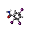


| #5: Chemical |  Mass: 498.826 Da / Num. of mol.: 3 / Source method: obtained synthetically / Formula: C7H4I3NO / Feature type: SUBJECT OF INVESTIGATION Mass: 498.826 Da / Num. of mol.: 3 / Source method: obtained synthetically / Formula: C7H4I3NO / Feature type: SUBJECT OF INVESTIGATION#7: Chemical |  Mass: 280.445 Da / Num. of mol.: 3 / Source method: obtained synthetically / Formula: C18H32O2 Mass: 280.445 Da / Num. of mol.: 3 / Source method: obtained synthetically / Formula: C18H32O2#8: Water | ChemComp-HOH / | Mass: 18.015 Da / Num. of mol.: 282 / Source method: isolated from a natural source / Formula: H2O |
|---|
-Details
| Has ligand of interest | Y |
|---|---|
| Has protein modification | Y |
-Experimental details
-Experiment
| Experiment | Method: ELECTRON MICROSCOPY |
|---|---|
| EM experiment | Aggregation state: PARTICLE / 3D reconstruction method: single particle reconstruction |
- Sample preparation
| Component | Name: SARS-CoV-2 Spike with ethylbenzamide-tri-iodo Siallyllactose, C3 symmetry Type: COMPLEX / Entity ID: #1 / Source: RECOMBINANT | ||||||||||||
|---|---|---|---|---|---|---|---|---|---|---|---|---|---|
| Source (natural) |
| ||||||||||||
| Source (recombinant) | Organism: Trichoplusia ni (cabbage looper) | ||||||||||||
| Buffer solution | pH: 7.5 | ||||||||||||
| Specimen | Embedding applied: NO / Shadowing applied: NO / Staining applied: NO / Vitrification applied: YES | ||||||||||||
| Vitrification | Cryogen name: ETHANE / Humidity: 100 % |
- Electron microscopy imaging
| Experimental equipment |  Model: Titan Krios / Image courtesy: FEI Company |
|---|---|
| Microscopy | Model: FEI TITAN KRIOS |
| Electron gun | Electron source:  FIELD EMISSION GUN / Accelerating voltage: 300 kV / Illumination mode: FLOOD BEAM FIELD EMISSION GUN / Accelerating voltage: 300 kV / Illumination mode: FLOOD BEAM |
| Electron lens | Mode: BRIGHT FIELD / Nominal defocus max: 2400 nm / Nominal defocus min: 800 nm |
| Image recording | Electron dose: 60 e/Å2 / Detector mode: COUNTING / Film or detector model: GATAN K2 SUMMIT (4k x 4k) |
- Processing
| Software |
| |||||||||
|---|---|---|---|---|---|---|---|---|---|---|
| EM software | Name: PHENIX / Category: model refinement | |||||||||
| CTF correction | Type: PHASE FLIPPING ONLY | |||||||||
| 3D reconstruction | Resolution: 2.27 Å / Resolution method: FSC 0.143 CUT-OFF / Num. of particles: 312018 / Symmetry type: POINT | |||||||||
| Refinement | Cross valid method: NONE Stereochemistry target values: GeoStd + Monomer Library + CDL v1.2 | |||||||||
| Displacement parameters | Biso mean: 55.43 Å2 |
