 Movie
Movie Controller


+ Open data

- Basic information
| Entry | Database: PDB / ID: 7qus | |||||||||||||||||||||||||||||||||
|---|---|---|---|---|---|---|---|---|---|---|---|---|---|---|---|---|---|---|---|---|---|---|---|---|---|---|---|---|---|---|---|---|---|---|
| Title | SARS-CoV-2 Spike, C3 symmetry | |||||||||||||||||||||||||||||||||
 Components Components | Spike glycoprotein,Fibritin | |||||||||||||||||||||||||||||||||
 Keywords Keywords | VIRAL PROTEIN / SARS-CoV-2 Spike | |||||||||||||||||||||||||||||||||
| Function / homology |  Function and homology information Function and homology informationvirion component / symbiont-mediated disruption of host tissue / Maturation of spike protein / Translation of Structural Proteins / Virion Assembly and Release / host cell surface / host extracellular region / symbiont-mediated-mediated suppression of host tetherin activity / Induction of Cell-Cell Fusion / structural constituent of virion ...virion component / symbiont-mediated disruption of host tissue / Maturation of spike protein / Translation of Structural Proteins / Virion Assembly and Release / host cell surface / host extracellular region / symbiont-mediated-mediated suppression of host tetherin activity / Induction of Cell-Cell Fusion / structural constituent of virion / positive regulation of viral entry into host cell / membrane fusion / host cell endoplasmic reticulum-Golgi intermediate compartment membrane / Attachment and Entry / entry receptor-mediated virion attachment to host cell / receptor-mediated virion attachment to host cell / host cell surface receptor binding / symbiont-mediated suppression of host innate immune response / endocytosis involved in viral entry into host cell / receptor ligand activity / fusion of virus membrane with host plasma membrane / fusion of virus membrane with host endosome membrane / viral envelope / symbiont entry into host cell / virion attachment to host cell / host cell plasma membrane / SARS-CoV-2 activates/modulates innate and adaptive immune responses / virion membrane / membrane / identical protein binding / plasma membrane Similarity search - Function | |||||||||||||||||||||||||||||||||
| Biological species |   Severe acute respiratory syndrome coronavirus 2Enterobacteria phage T4 (virus) Severe acute respiratory syndrome coronavirus 2Enterobacteria phage T4 (virus) | |||||||||||||||||||||||||||||||||
| Method | ELECTRON MICROSCOPY / single particle reconstruction / cryo EM / Resolution: 2.39 Å | |||||||||||||||||||||||||||||||||
 Authors Authors | Naismith, J.H. / Yang, Y. / Liu, J.W. | |||||||||||||||||||||||||||||||||
| Funding support | 1items
| |||||||||||||||||||||||||||||||||
 Citation Citation | Journal: Science / Year: 2022 Title: Pathogen-sugar interactions revealed by universal saturation transfer analysis. Authors: Charles J Buchanan / Ben Gaunt / Peter J Harrison / Yun Yang / Jiwei Liu / Aziz Khan / Andrew M Giltrap / Audrey Le Bas / Philip N Ward / Kapil Gupta / Maud Dumoux / Tiong Kit Tan / Lisa ...Authors: Charles J Buchanan / Ben Gaunt / Peter J Harrison / Yun Yang / Jiwei Liu / Aziz Khan / Andrew M Giltrap / Audrey Le Bas / Philip N Ward / Kapil Gupta / Maud Dumoux / Tiong Kit Tan / Lisa Schimaski / Sergio Daga / Nicola Picchiotti / Margherita Baldassarri / Elisa Benetti / Chiara Fallerini / Francesca Fava / Annarita Giliberti / Panagiotis I Koukos / Matthew J Davy / Abirami Lakshminarayanan / Xiaochao Xue / Georgios Papadakis / Lachlan P Deimel / Virgínia Casablancas-Antràs / Timothy D W Claridge / Alexandre M J J Bonvin / Quentin J Sattentau / Simone Furini / Marco Gori / Jiandong Huo / Raymond J Owens / Christiane Schaffitzel / Imre Berger / Alessandra Renieri / / James H Naismith / Andrew J Baldwin / Benjamin G Davis /     Abstract: Many pathogens exploit host cell-surface glycans. However, precise analyses of glycan ligands binding with heavily modified pathogen proteins can be confounded by overlapping sugar signals and/or ...Many pathogens exploit host cell-surface glycans. However, precise analyses of glycan ligands binding with heavily modified pathogen proteins can be confounded by overlapping sugar signals and/or compounded with known experimental constraints. Universal saturation transfer analysis (uSTA) builds on existing nuclear magnetic resonance spectroscopy to provide an automated workflow for quantitating protein-ligand interactions. uSTA reveals that early-pandemic, B-origin-lineage severe acute respiratory syndrome coronavirus 2 (SARS-CoV-2) spike trimer binds sialoside sugars in an "end-on" manner. uSTA-guided modeling and a high-resolution cryo-electron microscopy structure implicate the spike N-terminal domain (NTD) and confirm end-on binding. This finding rationalizes the effect of NTD mutations that abolish sugar binding in SARS-CoV-2 variants of concern. Together with genetic variance analyses in early pandemic patient cohorts, this binding implicates a sialylated polylactosamine motif found on tetraantennary N-linked glycoproteins deep in the human lung as potentially relevant to virulence and/or zoonosis. | |||||||||||||||||||||||||||||||||
| History |
|
- Structure visualization
| Structure viewer | Molecule: MolmilJmol/JSmol |
|---|
- Downloads & links
-Download
| PDBx/mmCIF format | 7qus.cif.gz | 821.9 KB | Display | PDBx/mmCIF format |
|---|---|---|---|---|
| PDB format | pdb7qus.ent.gz | 542.5 KB | Display | PDB format |
| PDBx/mmJSON format | 7qus.json.gz | Tree view | PDBx/mmJSON format | |
| Others |  Other downloads Other downloads |
-Validation report
| Arichive directory | https://data.pdbj.org/pub/pdb/validation_reports/qu/7qusftp://data.pdbj.org/pub/pdb/validation_reports/qu/7qus | HTTPS FTP |
|---|
-Related structure data
| Related structure data |  14153MC  7qurC M: map data used to model this data C: citing same article ( |
|---|---|
| Similar structure data |


-Links
 PDBj
PDBj


- Assembly
| Deposited unit | 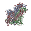
| ||||||||||||||||||||||||||||||||||||||||||||||||||||||||||||||||||||||||||||||||||||||||||||||||||||||||||||||||||||||||||||||||
|---|---|---|---|---|---|---|---|---|---|---|---|---|---|---|---|---|---|---|---|---|---|---|---|---|---|---|---|---|---|---|---|---|---|---|---|---|---|---|---|---|---|---|---|---|---|---|---|---|---|---|---|---|---|---|---|---|---|---|---|---|---|---|---|---|---|---|---|---|---|---|---|---|---|---|---|---|---|---|---|---|---|---|---|---|---|---|---|---|---|---|---|---|---|---|---|---|---|---|---|---|---|---|---|---|---|---|---|---|---|---|---|---|---|---|---|---|---|---|---|---|---|---|---|---|---|---|---|---|---|
| 1 |
| ||||||||||||||||||||||||||||||||||||||||||||||||||||||||||||||||||||||||||||||||||||||||||||||||||||||||||||||||||||||||||||||||
| Noncrystallographic symmetry (NCS) | NCS domain:
NCS domain segments: Ens-ID: ens_1
|