 Movie
Movie Controller

[English] 日本語
 Yorodumi
- PDB-7pwy: Structure of human dimeric ACMSD in complex with the inhibitor TE... -
+ Open data

- Basic information
| Entry | Database: PDB / ID: 7pwy | |||||||||
|---|---|---|---|---|---|---|---|---|---|---|
| Title | Structure of human dimeric ACMSD in complex with the inhibitor TES-1025 | |||||||||
 Components Components | 2-amino-3-carboxymuconate-6-semialdehyde decarboxylase | |||||||||
 Keywords Keywords | LYASE / ACMSD / de-novo NAD+ synthesis / TES-1025 / decarboxylase | |||||||||
| Function / homology |  Function and homology information Function and homology informationaminocarboxymuconate-semialdehyde decarboxylase activity / negative regulation of quinolinate biosynthetic process / picolinic acid biosynthetic process / regulation of 'de novo' NAD biosynthetic process from L-tryptophan / aminocarboxymuconate-semialdehyde decarboxylase / secondary metabolic process / L-tryptophan catabolic process / Tryptophan catabolism / hydrolase activity / zinc ion binding ...aminocarboxymuconate-semialdehyde decarboxylase activity / negative regulation of quinolinate biosynthetic process / picolinic acid biosynthetic process / regulation of 'de novo' NAD biosynthetic process from L-tryptophan / aminocarboxymuconate-semialdehyde decarboxylase / secondary metabolic process / L-tryptophan catabolic process / Tryptophan catabolism / hydrolase activity / zinc ion binding / cytosol / cytoplasm Similarity search - Function | |||||||||
| Biological species |  Homo sapiens (human) Homo sapiens (human) | |||||||||
| Method |  X-RAY DIFFRACTION / SYNCHROTRON / MOLECULAR REPLACEMENT / Resolution: 2.5 Å X-RAY DIFFRACTION / SYNCHROTRON / MOLECULAR REPLACEMENT / Resolution: 2.5 Å | |||||||||
 Authors Authors | Cianci, M. / Giacche, N. / Carotti, A. / Liscio, P. / Amici, A. / Cialabrini, L. / De Franco, F. / Pellicciari, R. / Raffaelli, N. | |||||||||
| Funding support | 1items
| |||||||||
 Citation Citation | Journal: Front Mol Biosci / Year: 2022 Title: Structural Basis of Human Dimeric alpha-Amino-beta-Carboxymuconate-epsilon-Semialdehyde Decarboxylase Inhibition With TES-1025. Authors: Cianci, M. / Giacche, N. / Cialabrini, L. / Carotti, A. / Liscio, P. / Rosatelli, E. / De Franco, F. / Gasparrini, M. / Robertson, J. / Amici, A. / Raffaelli, N. / Pellicciari, R. | |||||||||
| History |
|
- Structure visualization
| Structure viewer | Molecule: MolmilJmol/JSmol |
|---|
- Downloads & links
-Download
| PDBx/mmCIF format | 7pwy.cif.gz | 333.2 KB | Display | PDBx/mmCIF format |
|---|---|---|---|---|
| PDB format | pdb7pwy.ent.gz | 218.7 KB | Display | PDB format |
| PDBx/mmJSON format | 7pwy.json.gz | Tree view | PDBx/mmJSON format | |
| Others |  Other downloads Other downloads |
-Validation report
| Arichive directory | https://data.pdbj.org/pub/pdb/validation_reports/pw/7pwyftp://data.pdbj.org/pub/pdb/validation_reports/pw/7pwy | HTTPS FTP |
|---|
-Related structure data
| Related structure data | 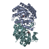 4ih3S S: Starting model for refinement |
|---|---|
| Similar structure data |
-Links
 PDBj
PDBj

- Assembly
| Deposited unit | 
| ||||||||||||
|---|---|---|---|---|---|---|---|---|---|---|---|---|---|
| 1 | 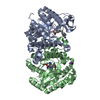
| ||||||||||||
| 2 | 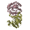
| ||||||||||||
| Unit cell |
| ||||||||||||
| Components on special symmetry positions |
|
-Components
| #1: Protein | Mass: 38084.223 Da / Num. of mol.: 4 Source method: isolated from a genetically manipulated source Source: (gene. exp.) Homo sapiens (human) / Gene: ACMSD / Production host:  Komagataella pastoris (fungus) Komagataella pastoris (fungus)References: UniProt: Q8TDX5, aminocarboxymuconate-semialdehyde decarboxylase #2: Chemical | ChemComp-ZN /   Mass: 65.409 Da / Num. of mol.: 4 / Source method: obtained synthetically / Formula: Zn Mass: 65.409 Da / Num. of mol.: 4 / Source method: obtained synthetically / Formula: Zn#3: Chemical | 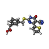  Mass: 383.444 Da / Num. of mol.: 2 / Source method: obtained synthetically / Formula: C18H13N3O3S2 / Feature type: SUBJECT OF INVESTIGATION Mass: 383.444 Da / Num. of mol.: 2 / Source method: obtained synthetically / Formula: C18H13N3O3S2 / Feature type: SUBJECT OF INVESTIGATION#4: Chemical | ChemComp-K /   Mass: 39.098 Da / Num. of mol.: 6 / Source method: isolated from a natural source / Formula: K Mass: 39.098 Da / Num. of mol.: 6 / Source method: isolated from a natural source / Formula: K#5: Water | ChemComp-HOH / |  Mass: 18.015 Da / Num. of mol.: 289 / Source method: isolated from a natural source / Formula: H2O Mass: 18.015 Da / Num. of mol.: 289 / Source method: isolated from a natural source / Formula: H2OHas ligand of interest | Y | |
|---|
-Experimental details
-Experiment
| Experiment | Method: X-RAY DIFFRACTION / Number of used crystals: 1 |
|---|
- Sample preparation
| Crystal | Density Matthews: 2.49 Å3/Da / Density % sol: 50.62 % |
|---|---|
| Crystal grow | Temperature: 291 K / Method: vapor diffusion, sitting drop / pH: 5.7 / Details: 100 mM Na(CH3COO), 22% (w/v) PEG 4000. |
-Data collection
| Diffraction | Mean temperature: 100 K / Serial crystal experiment: N |
|---|---|
| Diffraction source | Source: SYNCHROTRON / Site: ESRF  / Beamline: MASSIF-3 / Wavelength: 0.967 Å / Beamline: MASSIF-3 / Wavelength: 0.967 Å |
| Detector | Type: DECTRIS EIGER X 4M / Detector: PIXEL / Date: Sep 2, 2017 |
| Radiation | Monochromator: Si(111) / Protocol: SINGLE WAVELENGTH / Monochromatic (M) / Laue (L): M / Scattering type: x-ray |
| Radiation wavelength | Wavelength: 0.967 Å / Relative weight: 1 |
| Reflection | Resolution: 2.5→45.89 Å / Num. obs: 51945 / % possible obs: 99.9 % / Observed criterion σ(I): 1.6 / Redundancy: 7.6 % / Biso Wilson estimate: 52.35 Å2 / CC1/2: 0.999 / CC star: 1 / Rmerge(I) obs: 0.11 / Net I/σ(I): 11.5 |
| Reflection shell | Resolution: 2.5→2.58 Å / Redundancy: 6.8 % / Rmerge(I) obs: 1.1 / Mean I/σ(I) obs: 1.6 / Num. unique obs: 4423 / CC1/2: 0.766 / CC star: 0.926 / % possible all: 100 |
- Processing
| Software |
| |||||||||||||||||||||||||||||||||||||||||||||||||||||||||||||||||||||||||||||||||||||||||||||||||||||||||||||||||||||||||||||||||||||
|---|---|---|---|---|---|---|---|---|---|---|---|---|---|---|---|---|---|---|---|---|---|---|---|---|---|---|---|---|---|---|---|---|---|---|---|---|---|---|---|---|---|---|---|---|---|---|---|---|---|---|---|---|---|---|---|---|---|---|---|---|---|---|---|---|---|---|---|---|---|---|---|---|---|---|---|---|---|---|---|---|---|---|---|---|---|---|---|---|---|---|---|---|---|---|---|---|---|---|---|---|---|---|---|---|---|---|---|---|---|---|---|---|---|---|---|---|---|---|---|---|---|---|---|---|---|---|---|---|---|---|---|---|---|---|
| Refinement | Method to determine structure: MOLECULAR REPLACEMENT Starting model: 4IH3 Resolution: 2.5→45.33 Å / SU ML: 0.3606 / Cross valid method: FREE R-VALUE / σ(F): 1.33 / Phase error: 29.4096 Stereochemistry target values: GeoStd + Monomer Library + CDL v1.2
| |||||||||||||||||||||||||||||||||||||||||||||||||||||||||||||||||||||||||||||||||||||||||||||||||||||||||||||||||||||||||||||||||||||
| Solvent computation | Shrinkage radii: 0.9 Å / VDW probe radii: 1.11 Å / Solvent model: FLAT BULK SOLVENT MODEL | |||||||||||||||||||||||||||||||||||||||||||||||||||||||||||||||||||||||||||||||||||||||||||||||||||||||||||||||||||||||||||||||||||||
| Displacement parameters | Biso mean: 68.28 Å2 | |||||||||||||||||||||||||||||||||||||||||||||||||||||||||||||||||||||||||||||||||||||||||||||||||||||||||||||||||||||||||||||||||||||
| Refinement step | Cycle: LAST / Resolution: 2.5→45.33 Å
| |||||||||||||||||||||||||||||||||||||||||||||||||||||||||||||||||||||||||||||||||||||||||||||||||||||||||||||||||||||||||||||||||||||
| Refine LS restraints |
| |||||||||||||||||||||||||||||||||||||||||||||||||||||||||||||||||||||||||||||||||||||||||||||||||||||||||||||||||||||||||||||||||||||
| LS refinement shell |
|
