 Movie
Movie Controller

[English] 日本語
 Yorodumi
- PDB-7p8u: Crystal Structure of leukotoxin LukE from Staphylococcus aureus i... -
+ Open data

- Basic information
| Entry | Database: PDB / ID: 7p8u | ||||||
|---|---|---|---|---|---|---|---|
| Title | Crystal Structure of leukotoxin LukE from Staphylococcus aureus in complex with p-cresyl sulfate | ||||||
 Components Components | Leucotoxin LukEv | ||||||
 Keywords Keywords | TOXIN / leukotoxin / beta barrel pore forming toxin / cytolysis / hemolysis | ||||||
| Function / homology |  Function and homology information Function and homology information | ||||||
| Biological species |   Staphylococcus aureus (bacteria) Staphylococcus aureus (bacteria) | ||||||
| Method |  X-RAY DIFFRACTION / SYNCHROTRON / MOLECULAR REPLACEMENT / Resolution: 1.6 Å X-RAY DIFFRACTION / SYNCHROTRON / MOLECULAR REPLACEMENT / Resolution: 1.6 Å | ||||||
 Authors Authors | Lambey, P. / Hoh, F. / Peysson, F. / Granier, S. / Leyrat, C. | ||||||
| Funding support |  France, 1items France, 1items
| ||||||
 Citation Citation | Journal: Elife / Year: 2022 Title: Structural insights into recognition of chemokine receptors by Staphylococcus aureus leukotoxins. Authors: Lambey, P. / Otun, O. / Cong, X. / Hoh, F. / Brunel, L. / Verdie, P. / Grison, C.M. / Peysson, F. / Jeannot, S. / Durroux, T. / Bechara, C. / Granier, S. / Leyrat, C. | ||||||
| History |
|
- Structure visualization
| Structure viewer | Molecule: MolmilJmol/JSmol |
|---|
- Downloads & links
-Download
| PDBx/mmCIF format | 7p8u.cif.gz | 138.3 KB | Display | PDBx/mmCIF format |
|---|---|---|---|---|
| PDB format | pdb7p8u.ent.gz | 106.6 KB | Display | PDB format |
| PDBx/mmJSON format | 7p8u.json.gz | Tree view | PDBx/mmJSON format | |
| Others |  Other downloads Other downloads |
-Validation report
| Summary document | 7p8u_validation.pdf.gz | 1.3 MB | Display | wwPDB validaton report |
|---|---|---|---|---|
| Full document | 7p8u_full_validation.pdf.gz | 1.3 MB | Display | |
| Data in XML | 7p8u_validation.xml.gz | 15.5 KB | Display | |
| Data in CIF | 7p8u_validation.cif.gz | 23.2 KB | Display | |
| Arichive directory | https://data.pdbj.org/pub/pdb/validation_reports/p8/7p8uftp://data.pdbj.org/pub/pdb/validation_reports/p8/7p8u | HTTPS FTP |
-Related structure data
| Related structure data | 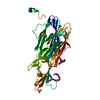 7p8sC 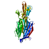 7p8tSC  7p8xC  7p93C S: Starting model for refinement C: citing same article ( |
|---|---|
| Similar structure data |
-Links
 PDBj
PDBj


- Assembly
| Deposited unit | 
| ||||||||
|---|---|---|---|---|---|---|---|---|---|
| 1 |
| ||||||||
| Unit cell |
|
-Components
-Protein , 1 types, 1 molecules A
| #1: Protein | Mass: 34686.691 Da / Num. of mol.: 1 Source method: isolated from a genetically manipulated source Source: (gene. exp.) Staphylococcus aureus (bacteria) / Gene: lukEv, SAOUHSC_01955 / Production host: |
|---|
-Non-polymers , 5 types, 266 molecules 


| #2: Chemical | ChemComp-PEG /  Mass: 106.120 Da / Num. of mol.: 1 / Source method: obtained synthetically / Formula: C4H10O3 Mass: 106.120 Da / Num. of mol.: 1 / Source method: obtained synthetically / Formula: C4H10O3 | ||
|---|---|---|---|
| #3: Chemical | ChemComp-IMD /  Mass: 69.085 Da / Num. of mol.: 1 / Source method: obtained synthetically / Formula: C3H5N2 Mass: 69.085 Da / Num. of mol.: 1 / Source method: obtained synthetically / Formula: C3H5N2 | ||
| #4: Chemical | ChemComp-SO4 /  Mass: 96.063 Da / Num. of mol.: 1 / Source method: obtained synthetically / Formula: SO4 Mass: 96.063 Da / Num. of mol.: 1 / Source method: obtained synthetically / Formula: SO4 | ||
| #5: Chemical |  Mass: 188.201 Da / Num. of mol.: 3 / Source method: obtained synthetically / Formula: C7H8O4S / Feature type: SUBJECT OF INVESTIGATION Mass: 188.201 Da / Num. of mol.: 3 / Source method: obtained synthetically / Formula: C7H8O4S / Feature type: SUBJECT OF INVESTIGATION#6: Water | ChemComp-HOH / | Mass: 18.015 Da / Num. of mol.: 260 / Source method: isolated from a natural source / Formula: H2O |
-Details
| Has ligand of interest | Y |
|---|
-Experimental details
-Experiment
| Experiment | Method: X-RAY DIFFRACTION / Number of used crystals: 1 |
|---|
- Sample preparation
| Crystal | Density Matthews: 2.63 Å3/Da / Density % sol: 53.23 % |
|---|---|
| Crystal grow | Temperature: 293.15 K / Method: vapor diffusion Details: 0.1 M Imidazole.HCl pH 8.0, 30% (w/v) MPD, 10% (w/v) PEG 4000 |
-Data collection
| Diffraction | Mean temperature: 100 K / Serial crystal experiment: N |
|---|---|
| Diffraction source | Source: SYNCHROTRON / Site: ESRF / Beamline: MASSIF-1 / Wavelength: 0.96545 Å |
| Detector | Type: DECTRIS PILATUS3 2M / Detector: PIXEL / Date: Jan 31, 2021 |
| Radiation | Protocol: SINGLE WAVELENGTH / Monochromatic (M) / Laue (L): M / Scattering type: x-ray |
| Radiation wavelength | Wavelength: 0.96545 Å / Relative weight: 1 |
| Reflection | Resolution: 1.6→47.65 Å / Num. obs: 48967 / % possible obs: 99.3 % / Redundancy: 3.4 % / CC1/2: 0.996 / Net I/σ(I): 15.5 |
| Reflection shell | Resolution: 1.6→1.63 Å / Num. unique obs: 2360 / CC1/2: 0.624 |
- Processing
| Software |
| ||||||||||||||||||||||||||||||||||||||||||||||||||||||||||||||||||
|---|---|---|---|---|---|---|---|---|---|---|---|---|---|---|---|---|---|---|---|---|---|---|---|---|---|---|---|---|---|---|---|---|---|---|---|---|---|---|---|---|---|---|---|---|---|---|---|---|---|---|---|---|---|---|---|---|---|---|---|---|---|---|---|---|---|---|---|
| Refinement | Method to determine structure: MOLECULAR REPLACEMENT Starting model: 7P8T Resolution: 1.6→47.65 Å / Cor.coef. Fo:Fc: 0.954 / Cor.coef. Fo:Fc free: 0.929 / SU R Cruickshank DPI: 0.077 / Cross valid method: THROUGHOUT / SU R Blow DPI: 0.08 / SU Rfree Blow DPI: 0.079 / SU Rfree Cruickshank DPI: 0.077
| ||||||||||||||||||||||||||||||||||||||||||||||||||||||||||||||||||
| Displacement parameters | Biso mean: 34.24 Å2
| ||||||||||||||||||||||||||||||||||||||||||||||||||||||||||||||||||
| Refine analyze | Luzzati coordinate error obs: 0.2 Å | ||||||||||||||||||||||||||||||||||||||||||||||||||||||||||||||||||
| Refinement step | Cycle: LAST / Resolution: 1.6→47.65 Å
| ||||||||||||||||||||||||||||||||||||||||||||||||||||||||||||||||||
| Refine LS restraints |
| ||||||||||||||||||||||||||||||||||||||||||||||||||||||||||||||||||
| LS refinement shell | Resolution: 1.6→1.61 Å
| ||||||||||||||||||||||||||||||||||||||||||||||||||||||||||||||||||
| Refinement TLS params. | Origin x: 14.7474 Å / Origin y: -14.4656 Å / Origin z: -12.3236 Å
| ||||||||||||||||||||||||||||||||||||||||||||||||||||||||||||||||||
| Refinement TLS group |
|
