- PDB-7p2u: Crystal structure of Schistosoma mansoni HDAC8 in complex with a ... -
+
データを開く
IDまたはキーワード:
読み込み中...
-
基本情報
登録情報
データベース: PDB / ID: 7p2u
タイトル
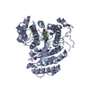
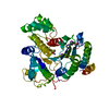
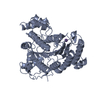
Crystal structure of Schistosoma mansoni HDAC8 in complex with a 3-chlorophenyl-spiroindoline capped hydroxamate-based inhibitor, bound to a novel site
根拠: Steady-state kinetics experiments confirmed that ligand behaved as an allosteric inhibitor; fluorescence spectra - at equilibrium - confirmed the binding at a novel binding site; all of the ...根拠: Steady-state kinetics experiments confirmed that ligand behaved as an allosteric inhibitor; fluorescence spectra - at equilibrium - confirmed the binding at a novel binding site; all of the above experiments were confirmed by dedicated mutant form. More details will be released in related publication
解像度: 1.8→46.63 Å / Cor.coef. Fo:Fc: 0.968 / Cor.coef. Fo:Fc free: 0.959 / SU B: 3.651 / SU ML: 0.103 / 交差検証法: THROUGHOUT / σ(F): 0 / ESU R: 0.116 / ESU R Free: 0.113 / 立体化学のターゲット値: MAXIMUM LIKELIHOOD 詳細: HYDROGENS HAVE BEEN ADDED IN THE RIDING POSITIONS U VALUES : REFINED INDIVIDUALLY. Used automatic weighting to optimize X-ray to stereochemistry weight
Rfactor
反射数
%反射
Selection details
Rfree
0.2132
2204
5 %
RANDOM
Rwork
0.179
-
-
-
obs
0.1808
42160
99.94 %
-
溶媒の処理
イオンプローブ半径: 0.8 Å / 減衰半径: 0.8 Å / VDWプローブ半径: 1.2 Å / 溶媒モデル: MASK
 ムービー
ムービー コントローラー


 データを開く
 基本情報
基本情報 要素
要素 キーワード
キーワード 機能・相同性情報
 X線回折 /
  データ登録者
 引用
引用 構造の表示
構造の表示 ダウンロードとリンク
 その他のダウンロード

 PDBj
PDBj
 集合体
集合体


 分子量: 65.409 Da / 分子数: 1 / 由来タイプ: 合成 / 式: Zn
 分子量: 39.098 Da / 分子数: 3 / 由来タイプ: 合成 / 式: K
 分子量: 35.453 Da / 分子数: 1 / 由来タイプ: 合成 / 式: Cl
 分子量: 150.087 Da / 分子数: 1 / 由来タイプ: 合成 / 式: C4H6O6
 分子量: 468.011 Da / 分子数: 1 / 由来タイプ: 合成 / 式: C25H26ClN3O2S / タイプ: SUBJECT OF INVESTIGATION
 試料調製
試料調製 / ビームライン: 11.2C / 波長: 1 Å
 解析
解析