 Movie
Movie Controller

[English] 日本語
 Yorodumi
- PDB-7p2t: Tetartohedrally twinned crystal structure of Schistosoma mansoni ... -
+ Open data

- Basic information
| Entry | Database: PDB / ID: 7p2t | ||||||
|---|---|---|---|---|---|---|---|
| Title | Tetartohedrally twinned crystal structure of Schistosoma mansoni HDAC8 in complex with a tricyclic thieno[3,2-b]indole capped hydroxamate-based inhibitor, bromine derivative | ||||||
 Components Components | Histone deacetylase 8 | ||||||
 Keywords Keywords | HYDROLASE / SmHDAC8 / active-site / HDACi / histone-deacetilase inhibitor complex / tetartohedral / twinning | ||||||
| Function / homology |  Function and homology information Function and homology informationhistone deacetylase activity, hydrolytic mechanism / histone deacetylase / heterochromatin formation / metal ion binding / nucleus Similarity search - Function | ||||||
| Biological species |  | ||||||
| Method |  X-RAY DIFFRACTION / SYNCHROTRON / MOLECULAR REPLACEMENT / Resolution: 2.3 Å X-RAY DIFFRACTION / SYNCHROTRON / MOLECULAR REPLACEMENT / Resolution: 2.3 Å | ||||||
 Authors Authors | Saccoccia, F. / Gemma, S. / Campiani, G. / Ruberti, G. | ||||||
 Citation Citation | Journal: J.Biol.Chem. / Year: 2022 Title: Crystal structures of Schistosoma mansoni histone deacetylase 8 reveal a novel binding site for allosteric inhibitors. Authors: Saccoccia, F. / Pozzetti, L. / Gimmelli, R. / Butini, S. / Guidi, A. / Papoff, G. / Giannaccari, M. / Brogi, S. / Scognamiglio, V. / Gemma, S. / Ruberti, G. / Campiani, G. | ||||||
| History |
|
- Structure visualization
| Structure viewer | Molecule: MolmilJmol/JSmol |
|---|
- Downloads & links
-Download
| PDBx/mmCIF format | 7p2t.cif.gz | 318.6 KB | Display | PDBx/mmCIF format |
|---|---|---|---|---|
| PDB format | pdb7p2t.ent.gz | 257.5 KB | Display | PDB format |
| PDBx/mmJSON format | 7p2t.json.gz | Tree view | PDBx/mmJSON format | |
| Others |  Other downloads Other downloads |
-Validation report
| Arichive directory | https://data.pdbj.org/pub/pdb/validation_reports/p2/7p2tftp://data.pdbj.org/pub/pdb/validation_reports/p2/7p2t | HTTPS FTP |
|---|
-Related structure data
| Related structure data |  7p2sC  7p2uC  7p2vC 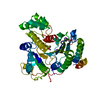 7pozC 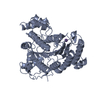 4bz5S S: Starting model for refinement C: citing same article ( |
|---|---|
| Similar structure data |
-Links
 PDBj
PDBj- Assembly
| Deposited unit | 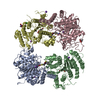
| ||||||||
|---|---|---|---|---|---|---|---|---|---|
| 1 | 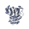
| ||||||||
| 2 | 
| ||||||||
| 3 | 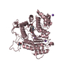
| ||||||||
| 4 | 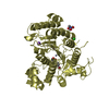
| ||||||||
| Unit cell |
|
-Components
-Protein , 1 types, 4 molecules ABCD
| #1: Protein | Mass: 49834.219 Da / Num. of mol.: 4 Source method: isolated from a genetically manipulated source Source: (gene. exp.)  |
|---|
-Non-polymers , 6 types, 83 molecules 


| #2: Chemical | ChemComp-ZN /  Mass: 65.409 Da / Num. of mol.: 4 / Source method: obtained synthetically / Formula: Zn Mass: 65.409 Da / Num. of mol.: 4 / Source method: obtained synthetically / Formula: Zn#3: Chemical | ChemComp-K /  Mass: 39.098 Da / Num. of mol.: 10 / Source method: obtained synthetically / Formula: K Mass: 39.098 Da / Num. of mol.: 10 / Source method: obtained synthetically / Formula: K#4: Chemical | ChemComp-4VX /  Mass: 485.417 Da / Num. of mol.: 4 / Source method: obtained synthetically / Formula: C22H17BrN2O2S2 / Feature type: SUBJECT OF INVESTIGATION Mass: 485.417 Da / Num. of mol.: 4 / Source method: obtained synthetically / Formula: C22H17BrN2O2S2 / Feature type: SUBJECT OF INVESTIGATION#5: Chemical |  Mass: 122.143 Da / Num. of mol.: 2 / Source method: obtained synthetically / Formula: C4H12NO3 Mass: 122.143 Da / Num. of mol.: 2 / Source method: obtained synthetically / Formula: C4H12NO3#6: Chemical | ChemComp-CL / |  Mass: 35.453 Da / Num. of mol.: 1 / Source method: obtained synthetically / Formula: Cl Mass: 35.453 Da / Num. of mol.: 1 / Source method: obtained synthetically / Formula: Cl#7: Water | ChemComp-HOH / | Mass: 18.015 Da / Num. of mol.: 62 / Source method: isolated from a natural source / Formula: H2O |
|---|
-Details
| Has ligand of interest | Y |
|---|
-Experimental details
-Experiment
| Experiment | Method: X-RAY DIFFRACTION / Number of used crystals: 1 |
|---|
- Sample preparation
| Crystal | Density Matthews: 2.19 Å3/Da / Density % sol: 43.92 % |
|---|---|
| Crystal grow | Temperature: 277.15 K / Method: vapor diffusion, hanging drop / Details: 20-22% PEG 3350, 200mM sodium/potassium tartrate / Temp details: cold room |
-Data collection
| Diffraction | Mean temperature: 100 K / Serial crystal experiment: N | |||||||||||||||||||||||||||||||||||||||||||||||||||||||||||||||||||||||||||||||||||||||||||||||||||||||||||||||||||||||||
|---|---|---|---|---|---|---|---|---|---|---|---|---|---|---|---|---|---|---|---|---|---|---|---|---|---|---|---|---|---|---|---|---|---|---|---|---|---|---|---|---|---|---|---|---|---|---|---|---|---|---|---|---|---|---|---|---|---|---|---|---|---|---|---|---|---|---|---|---|---|---|---|---|---|---|---|---|---|---|---|---|---|---|---|---|---|---|---|---|---|---|---|---|---|---|---|---|---|---|---|---|---|---|---|---|---|---|---|---|---|---|---|---|---|---|---|---|---|---|---|---|---|---|
| Diffraction source | Source: SYNCHROTRON / Site: ELETTRA  / Beamline: 11.2C / Wavelength: 1 Å / Beamline: 11.2C / Wavelength: 1 Å | |||||||||||||||||||||||||||||||||||||||||||||||||||||||||||||||||||||||||||||||||||||||||||||||||||||||||||||||||||||||||
| Detector | Type: DECTRIS PILATUS 6M / Detector: PIXEL / Date: Aug 4, 2020 | |||||||||||||||||||||||||||||||||||||||||||||||||||||||||||||||||||||||||||||||||||||||||||||||||||||||||||||||||||||||||
| Radiation | Monochromator: Double Crystal Si111 with LN2 closed loop cooling Protocol: SINGLE WAVELENGTH / Monochromatic (M) / Laue (L): M / Scattering type: x-ray | |||||||||||||||||||||||||||||||||||||||||||||||||||||||||||||||||||||||||||||||||||||||||||||||||||||||||||||||||||||||||
| Radiation wavelength | Wavelength: 1 Å / Relative weight: 1 | |||||||||||||||||||||||||||||||||||||||||||||||||||||||||||||||||||||||||||||||||||||||||||||||||||||||||||||||||||||||||
| Reflection twin |
| |||||||||||||||||||||||||||||||||||||||||||||||||||||||||||||||||||||||||||||||||||||||||||||||||||||||||||||||||||||||||
| Reflection | Resolution: 2.3→178.539 Å / Num. all: 76754 / Num. obs: 76754 / % possible obs: 100 % / Redundancy: 6.5 % / Rpim(I) all: 0.087 / Rrim(I) all: 0.226 / Rsym value: 0.208 / Net I/av σ(I): 2.3 / Net I/σ(I): 4.6 / Num. measured all: 501220 | |||||||||||||||||||||||||||||||||||||||||||||||||||||||||||||||||||||||||||||||||||||||||||||||||||||||||||||||||||||||||
| Reflection shell | Diffraction-ID: 1
|
- Processing
| Software |
| ||||||||||||||||||
|---|---|---|---|---|---|---|---|---|---|---|---|---|---|---|---|---|---|---|---|
| Refinement | Method to determine structure: MOLECULAR REPLACEMENT Starting model: 4bz5 Resolution: 2.3→48.063 Å / Cross valid method: THROUGHOUT
| ||||||||||||||||||
| Displacement parameters | Biso max: 119.88 Å2 / Biso mean: 50.183 Å2 / Biso min: 20.72 Å2 | ||||||||||||||||||
| Refinement step | Cycle: LAST / Resolution: 2.3→48.063 Å
|
