 Movie
Movie Controller

[English] 日本語
 Yorodumi
- PDB-7ok3: Crystal Structure of KRasG13C in Complex with Nucleotide-based Co... -
+ Open data

- Basic information
| Entry | Database: PDB / ID: 7ok3 | |||||||||
|---|---|---|---|---|---|---|---|---|---|---|
| Title | Crystal Structure of KRasG13C in Complex with Nucleotide-based Covalent Inhibitor edaGDP | |||||||||
 Components Components | Isoform 2B of GTPase KRas | |||||||||
 Keywords Keywords | HYDROLASE / GTPase / Ras / KRas / KRasG13C / Nucleotide analogues / edaGDP | |||||||||
| Function / homology | small monomeric GTPase / Ca2+ pathway / P-loop containing nucleotide triphosphate hydrolases / Rossmann fold / 3-Layer(aba) Sandwich / Alpha Beta / edaGDP / Isoform 2B of GTPase KRas Function and homology information Function and homology information | |||||||||
| Biological species |  Homo sapiens (human) Homo sapiens (human) | |||||||||
| Method |  X-RAY DIFFRACTION / SYNCHROTRON / MOLECULAR REPLACEMENT / Resolution: 1.6 Å X-RAY DIFFRACTION / SYNCHROTRON / MOLECULAR REPLACEMENT / Resolution: 1.6 Å | |||||||||
 Authors Authors | Goebel, L. / Mueller, M.P. / Rauh, D. | |||||||||
| Funding support |  Germany, 2items Germany, 2items
| |||||||||
 Citation Citation | Journal: Elife / Year: 2023 Title: Targeting oncogenic KRasG13C with nucleotide-based covalent inhibitors. Authors: Goebel, L. / Kirschner, T. / Koska, S. / Rai, A. / Janning, P. / Maffini, S. / Vatheuer, H. / Czodrowski, P. / Goody, R.S. / Muller, M.P. / Rauh, D. | |||||||||
| History |
|
- Structure visualization
| Structure viewer | Molecule: MolmilJmol/JSmol |
|---|
- Downloads & links
-Download
| PDBx/mmCIF format | 7ok3.cif.gz | 94.2 KB | Display | PDBx/mmCIF format |
|---|---|---|---|---|
| PDB format | pdb7ok3.ent.gz | 70.4 KB | Display | PDB format |
| PDBx/mmJSON format | 7ok3.json.gz | Tree view | PDBx/mmJSON format | |
| Others |  Other downloads Other downloads |
-Validation report
| Arichive directory | https://data.pdbj.org/pub/pdb/validation_reports/ok/7ok3ftp://data.pdbj.org/pub/pdb/validation_reports/ok/7ok3 | HTTPS FTP |
|---|
-Related structure data
| Related structure data |  7ok4C 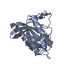 4obeS S: Starting model for refinement C: citing same article ( |
|---|---|
| Similar structure data |
-Links
 PDBj
PDBj


- Assembly
| Deposited unit | 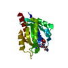
| ||||||||
|---|---|---|---|---|---|---|---|---|---|
| 1 |
| ||||||||
| Unit cell |
|
-Components
| #1: Protein | Mass: 19433.881 Da / Num. of mol.: 1 / Mutation: G13C Source method: isolated from a genetically manipulated source Source: (gene. exp.) Homo sapiens (human) / Gene: KRAS, KRAS2, RASK2 / Production host:  |
|---|---|
| #2: Chemical | ChemComp-VJ8 /   Mass: 585.356 Da / Num. of mol.: 1 / Source method: obtained synthetically / Formula: C16H25N7O13P2 / Feature type: SUBJECT OF INVESTIGATION Mass: 585.356 Da / Num. of mol.: 1 / Source method: obtained synthetically / Formula: C16H25N7O13P2 / Feature type: SUBJECT OF INVESTIGATION |
| #3: Water | ChemComp-HOH /  Mass: 18.015 Da / Num. of mol.: 187 / Source method: isolated from a natural source / Formula: H2O Mass: 18.015 Da / Num. of mol.: 187 / Source method: isolated from a natural source / Formula: H2O |
| Has ligand of interest | Y |
| Has protein modification | Y |
-Experimental details
-Experiment
| Experiment | Method: X-RAY DIFFRACTION / Number of used crystals: 1 |
|---|
- Sample preparation
| Crystal | Density Matthews: 2.17 Å3/Da / Density % sol: 43.29 % |
|---|---|
| Crystal grow | Temperature: 293 K / Method: vapor diffusion, hanging drop Details: 0.2 M (NH4)F, 20 % PEG3350, 67 mg/mL KRasG13C-edaGDP, 0.1 uL reservoir + 0.1 uL protein solution |
-Data collection
| Diffraction | Mean temperature: 100 K / Serial crystal experiment: N |
|---|---|
| Diffraction source | Source: SYNCHROTRON / Site: SLS  / Beamline: X10SA / Wavelength: 0.915 Å / Beamline: X10SA / Wavelength: 0.915 Å |
| Detector | Type: DECTRIS PILATUS 6M / Detector: PIXEL / Date: Sep 24, 2018 |
| Radiation | Protocol: SINGLE WAVELENGTH / Monochromatic (M) / Laue (L): M / Scattering type: x-ray |
| Radiation wavelength | Wavelength: 0.915 Å / Relative weight: 1 |
| Reflection | Resolution: 1.6→41.243 Å / Num. obs: 22009 / % possible obs: 100 % / Redundancy: 10.1 % / CC1/2: 0.99 / Rmerge(I) obs: 0.098 / Rrim(I) all: 0.103 / Net I/σ(I): 12.71 |
| Reflection shell | Resolution: 1.6→1.7 Å / Rmerge(I) obs: 0.966 / Num. unique obs: 3625 / CC1/2: 0.801 / Rrim(I) all: 1.016 / % possible all: 100 |
- Processing
| Software |
| ||||||||||||||||||||||||||||||||||||||||||||||||||||||
|---|---|---|---|---|---|---|---|---|---|---|---|---|---|---|---|---|---|---|---|---|---|---|---|---|---|---|---|---|---|---|---|---|---|---|---|---|---|---|---|---|---|---|---|---|---|---|---|---|---|---|---|---|---|---|---|
| Refinement | Method to determine structure: MOLECULAR REPLACEMENT Starting model: 4OBE Resolution: 1.6→41.24 Å / SU ML: 0.2 / Cross valid method: THROUGHOUT / σ(F): 1.38 / Phase error: 21.09 / Stereochemistry target values: ML
| ||||||||||||||||||||||||||||||||||||||||||||||||||||||
| Solvent computation | Shrinkage radii: 0.9 Å / VDW probe radii: 1.11 Å / Solvent model: FLAT BULK SOLVENT MODEL | ||||||||||||||||||||||||||||||||||||||||||||||||||||||
| Displacement parameters | Biso max: 83.76 Å2 / Biso mean: 27.867 Å2 / Biso min: 13.33 Å2 | ||||||||||||||||||||||||||||||||||||||||||||||||||||||
| Refinement step | Cycle: final / Resolution: 1.6→41.24 Å
| ||||||||||||||||||||||||||||||||||||||||||||||||||||||
| LS refinement shell | Refine-ID: X-RAY DIFFRACTION / Rfactor Rfree error: 0 / Total num. of bins used: 8 / % reflection obs: 100 %
| ||||||||||||||||||||||||||||||||||||||||||||||||||||||
| Refinement TLS params. | Method: refined / Origin x: 55.4797 Å / Origin y: 20.7789 Å / Origin z: 12.1359 Å
| ||||||||||||||||||||||||||||||||||||||||||||||||||||||
| Refinement TLS group |
|
