 Movie
Movie Controller

[English] 日本語
 Yorodumi
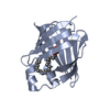
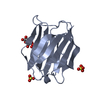
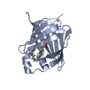
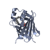
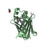
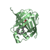
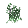
- PDB-7jx2: Cellular retinol-binding protein 2 (CRBP2) in complex with 2-palm... -
+ Open data

- Basic information
| Entry | Database: PDB / ID: 7jx2 | |||||||||
|---|---|---|---|---|---|---|---|---|---|---|
| Title | Cellular retinol-binding protein 2 (CRBP2) in complex with 2-palmitoylglycerol | |||||||||
 Components Components | Retinol-binding protein 2 | |||||||||
 Keywords Keywords | LIPID BINDING PROTEIN / palmitoylglycerol / monoacylglycerol / retinol-binding protein / lipid binding | |||||||||
| Function / homology |  Function and homology information Function and homology informationvitamin A metabolic process / triglyceride biosynthetic process / retinoid binding / retinal binding / molecular carrier activity / retinol binding / epidermis development / fatty acid transport / Retinoid metabolism and transport / fatty acid binding ...vitamin A metabolic process / triglyceride biosynthetic process / retinoid binding / retinal binding / molecular carrier activity / retinol binding / epidermis development / fatty acid transport / Retinoid metabolism and transport / fatty acid binding / nucleus / cytosol Similarity search - Function | |||||||||
| Biological species |  Homo sapiens (human) Homo sapiens (human) | |||||||||
| Method |  X-RAY DIFFRACTION / SYNCHROTRON / MOLECULAR REPLACEMENT / Resolution: 1.8 Å X-RAY DIFFRACTION / SYNCHROTRON / MOLECULAR REPLACEMENT / Resolution: 1.8 Å | |||||||||
 Authors Authors | Silvaroli, J.A. / Banarjee, S. / Golczak, M. | |||||||||
| Funding support |  United States, 2items United States, 2items
| |||||||||
 Citation Citation | Journal: J.Lipid Res. / Year: 2021 Title: Molecular basis for the interaction of cellular retinol binding protein 2 (CRBP2) with nonretinoid ligands. Authors: Silvaroli, J.A. / Plau, J. / Adams, C.H. / Banerjee, S. / Widjaja-Adhi, M.A.K. / Blaner, W.S. / Golczak, M. | |||||||||
| History |
|
- Structure visualization
| Structure viewer | Molecule: MolmilJmol/JSmol |
|---|
- Downloads & links
-Download
| PDBx/mmCIF format | 7jx2.cif.gz | 83.3 KB | Display | PDBx/mmCIF format |
|---|---|---|---|---|
| PDB format | pdb7jx2.ent.gz | 60 KB | Display | PDB format |
| PDBx/mmJSON format | 7jx2.json.gz | Tree view | PDBx/mmJSON format | |
| Others |  Other downloads Other downloads |
-Validation report
| Arichive directory | https://data.pdbj.org/pub/pdb/validation_reports/jx/7jx2ftp://data.pdbj.org/pub/pdb/validation_reports/jx/7jx2 | HTTPS FTP |
|---|
-Related structure data
| Related structure data |  7jvgC  7jvyC  7jwdC  7jwrC  7jz5C  7k3iC  6btiS S: Starting model for refinement C: citing same article ( |
|---|---|
| Similar structure data |


-Links
 PDBj
PDBj


- Assembly
| Deposited unit | 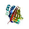
| ||||||||
|---|---|---|---|---|---|---|---|---|---|
| 1 |
| ||||||||
| Unit cell |
|
-Components
| #1: Protein | Mass: 16195.246 Da / Num. of mol.: 1 Source method: isolated from a genetically manipulated source Source: (gene. exp.) Homo sapiens (human) / Gene: RBP2, CRBP2 / Production host:  |
|---|---|
| #2: Chemical | ChemComp-VLP /   Mass: 330.503 Da / Num. of mol.: 1 / Source method: obtained synthetically / Formula: C19H38O4 / Feature type: SUBJECT OF INVESTIGATION Mass: 330.503 Da / Num. of mol.: 1 / Source method: obtained synthetically / Formula: C19H38O4 / Feature type: SUBJECT OF INVESTIGATION |
| #3: Water | ChemComp-HOH /  Mass: 18.015 Da / Num. of mol.: 209 / Source method: isolated from a natural source / Formula: H2O Mass: 18.015 Da / Num. of mol.: 209 / Source method: isolated from a natural source / Formula: H2O |
| Has ligand of interest | Y |
-Experimental details
-Experiment
| Experiment | Method: X-RAY DIFFRACTION / Number of used crystals: 1 |
|---|
- Sample preparation
| Crystal | Density Matthews: 2.51 Å3/Da / Density % sol: 51.09 % |
|---|---|
| Crystal grow | Temperature: 293 K / Method: vapor diffusion, sitting drop / pH: 8 / Details: 0.1 M Tris HCl, PEG 3350 22-28% |
-Data collection
| Diffraction | Mean temperature: 80 K / Serial crystal experiment: N |
|---|---|
| Diffraction source | Source: SYNCHROTRON / Site: APS / Beamline: 24-ID-C / Wavelength: 0.9791 Å |
| Detector | Type: DECTRIS PILATUS 6M-F / Detector: PIXEL / Date: Mar 13, 2018 |
| Radiation | Protocol: SINGLE WAVELENGTH / Monochromatic (M) / Laue (L): M / Scattering type: x-ray |
| Radiation wavelength | Wavelength: 0.9791 Å / Relative weight: 1 |
| Reflection | Resolution: 1.8→66.58 Å / Num. obs: 15466 / % possible obs: 99.7 % / Redundancy: 6.3 % / CC1/2: 0.996 / Rmerge(I) obs: 0.113 / Rpim(I) all: 0.072 / Rrim(I) all: 0.134 / Net I/σ(I): 8.2 |
| Reflection shell | Resolution: 1.8→1.84 Å / Redundancy: 6.6 % / Rmerge(I) obs: 0.873 / Mean I/σ(I) obs: 2.3 / Num. unique obs: 889 / CC1/2: 0.881 / Rpim(I) all: 0.554 / % possible all: 100 |
- Processing
| Software |
| ||||||||||||||||||||||||||||||||||||||||||
|---|---|---|---|---|---|---|---|---|---|---|---|---|---|---|---|---|---|---|---|---|---|---|---|---|---|---|---|---|---|---|---|---|---|---|---|---|---|---|---|---|---|---|---|
| Refinement | Method to determine structure: MOLECULAR REPLACEMENT Starting model: 6BTI Resolution: 1.8→46.31 Å / SU ML: 0.2 / Cross valid method: FREE R-VALUE / σ(F): 1.34 / Phase error: 29.44 / Stereochemistry target values: ML
| ||||||||||||||||||||||||||||||||||||||||||
| Solvent computation | Shrinkage radii: 0.9 Å / VDW probe radii: 1.11 Å / Solvent model: FLAT BULK SOLVENT MODEL | ||||||||||||||||||||||||||||||||||||||||||
| Refinement step | Cycle: LAST / Resolution: 1.8→46.31 Å
| ||||||||||||||||||||||||||||||||||||||||||
| Refine LS restraints |
| ||||||||||||||||||||||||||||||||||||||||||
| LS refinement shell |
| ||||||||||||||||||||||||||||||||||||||||||
| Refinement TLS params. | Method: refined / Origin x: -4.0167 Å / Origin y: -10.3064 Å / Origin z: 15.5622 Å
| ||||||||||||||||||||||||||||||||||||||||||
| Refinement TLS group | Selection details: all |
