 ムービー
ムービー コントローラー


+ データを開く

- 基本情報
基本情報
| 登録情報 | データベース: PDB / ID: 7jfy | ||||||
|---|---|---|---|---|---|---|---|
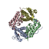
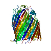
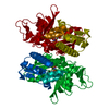
| タイトル | GAS41 YEATS domain in complex with 5 | ||||||
 要素 要素 | YEATS domain-containing protein 4 | ||||||
 キーワード キーワード | PROTEIN BINDING / Protein-inhibitor complex | ||||||
| 機能・相同性 |  機能・相同性情報 機能・相同性情報histone H3K18ac reader activity / histone H3K27ac reader activity / Activation of the TFAP2 (AP-2) family of transcription factors / regulation of double-strand break repair / NuA4 histone acetyltransferase complex / positive regulation of double-strand break repair via homologous recombination / structural constituent of cytoskeleton / nuclear matrix / nucleosome / mitotic cell cycle ...histone H3K18ac reader activity / histone H3K27ac reader activity / Activation of the TFAP2 (AP-2) family of transcription factors / regulation of double-strand break repair / NuA4 histone acetyltransferase complex / positive regulation of double-strand break repair via homologous recombination / structural constituent of cytoskeleton / nuclear matrix / nucleosome / mitotic cell cycle / HATs acetylate histones / histone binding / nuclear membrane / regulation of apoptotic process / regulation of cell cycle / chromatin remodeling / regulation of transcription by RNA polymerase II / regulation of DNA-templated transcription / positive regulation of DNA-templated transcription / nucleoplasm / nucleus 類似検索 - 分子機能 | ||||||
| 生物種 |  Homo sapiens (ヒト) Homo sapiens (ヒト) | ||||||
| 手法 |  X線回折 / シンクロトロン / 分子置換 / 解像度: 2.10055713817 Å X線回折 / シンクロトロン / 分子置換 / 解像度: 2.10055713817 Å | ||||||
 データ登録者 データ登録者 | Linhares, B.M. / Listunov, D. / Winkler, A. / Grembecka, J. / Cierpicki, T. | ||||||
| 資金援助 |  米国, 1件 米国, 1件
| ||||||
 引用 引用 | ジャーナル: To Be Published タイトル: GAS41 YEATS domain in complex with 5 著者: Linhares, B.M. / Listunov, D. / Winkler, A. / Grembecka, J. / Cierpicki, T. | ||||||
| 履歴 |
|
- 構造の表示
構造の表示
| 構造ビューア | 分子: MolmilJmol/JSmol |
|---|
- ダウンロードとリンク
-ダウンロード
| PDBx/mmCIF形式 | 7jfy.cif.gz | 167.1 KB | 表示 | PDBx/mmCIF形式 |
|---|---|---|---|---|
| PDB形式 | pdb7jfy.ent.gz | 104.5 KB | 表示 | PDB形式 |
| PDBx/mmJSON形式 | 7jfy.json.gz | ツリー表示 | PDBx/mmJSON形式 | |
| その他 |  その他のダウンロード その他のダウンロード |
-検証レポート
| 文書・要旨 | 7jfy_validation.pdf.gz | 1.4 MB | 表示 | wwPDB検証レポート |
|---|---|---|---|---|
| 文書・詳細版 | 7jfy_full_validation.pdf.gz | 1.4 MB | 表示 | |
| XML形式データ | 7jfy_validation.xml.gz | 31.2 KB | 表示 | |
| CIF形式データ | 7jfy_validation.cif.gz | 44.3 KB | 表示 | |
| アーカイブディレクトリ | https://data.pdbj.org/pub/pdb/validation_reports/jf/7jfyftp://data.pdbj.org/pub/pdb/validation_reports/jf/7jfy | HTTPS FTP |
-関連構造データ
| 関連構造データ | 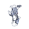 5vnaS S: 精密化の開始モデル |
|---|---|
| 類似構造データ |


-リンク
 PDBj
PDBj
- 集合体
集合体
| 登録構造単位 | 
| ||||||||||||||||||||||||
|---|---|---|---|---|---|---|---|---|---|---|---|---|---|---|---|---|---|---|---|---|---|---|---|---|---|
| 1 |
| ||||||||||||||||||||||||
| 単位格子 |
| ||||||||||||||||||||||||
| Components on special symmetry positions |
|
-要素
| #1: タンパク質 | 分子量: 17802.453 Da / 分子数: 4 / 由来タイプ: 組換発現 / 由来: (組換発現) Homo sapiens (ヒト) / 遺伝子: YEATS4, GAS41 / 発現宿主:  #2: 化合物 | ChemComp-V91 /   分子量: 362.470 Da / 分子数: 4 / 由来タイプ: 合成 / 式: C16H18N4O2S2 / タイプ: SUBJECT OF INVESTIGATION 分子量: 362.470 Da / 分子数: 4 / 由来タイプ: 合成 / 式: C16H18N4O2S2 / タイプ: SUBJECT OF INVESTIGATION#3: 化合物 | ChemComp-EDO /   分子量: 62.068 Da / 分子数: 6 / 由来タイプ: 合成 / 式: C2H6O2 分子量: 62.068 Da / 分子数: 6 / 由来タイプ: 合成 / 式: C2H6O2#4: 化合物 | ChemComp-DMS / |   分子量: 78.133 Da / 分子数: 1 / 由来タイプ: 合成 / 式: C2H6OS / コメント: DMSO, 沈殿剤*YM 分子量: 78.133 Da / 分子数: 1 / 由来タイプ: 合成 / 式: C2H6OS / コメント: DMSO, 沈殿剤*YM#5: 水 | ChemComp-HOH / |  分子量: 18.015 Da / 分子数: 728 / 由来タイプ: 天然 / 式: H2O 分子量: 18.015 Da / 分子数: 728 / 由来タイプ: 天然 / 式: H2O研究の焦点であるリガンドがあるか | Y | |
|---|
-実験情報
-実験
| 実験 | 手法: X線回折 / 使用した結晶の数: 1 |
|---|
- 試料調製
試料調製
| 結晶 | マシュー密度: 2.62 Å3/Da / 溶媒含有率: 52.96 % |
|---|---|
| 結晶化 | 温度: 277 K / 手法: 蒸気拡散法, シッティングドロップ法 / pH: 7.3 詳細: 0.1 M Tris, pH 7.3, 0.2 M magnesium chloride, 2.2 M sodium chloride |
-データ収集
| 回折 | 平均測定温度: 100 K / Serial crystal experiment: N |
|---|---|
| 放射光源 | 由来: シンクロトロン / サイト: APS / ビームライン: 21-ID-F / 波長: 0.97857 Å |
| 検出器 | タイプ: MARMOSAIC 325 mm CCD / 検出器: CCD / 日付: 2018年10月13日 |
| 放射 | プロトコル: SINGLE WAVELENGTH / 単色(M)・ラウエ(L): M / 散乱光タイプ: x-ray |
| 放射波長 | 波長: 0.97857 Å / 相対比: 1 |
| 反射 | 解像度: 2.1→39.85 Å / Num. obs: 42637 / % possible obs: 98.13 % / 冗長度: 3.9 % / Biso Wilson estimate: 30.2473095888 Å2 / CC1/2: 0.994 / CC star: 0.999 / Rmerge(I) obs: 0.09119 / Rpim(I) all: 0.05365 / Rrim(I) all: 0.1059 / Net I/σ(I): 11.74 |
| 反射 シェル | 解像度: 2.101→2.176 Å / Rmerge(I) obs: 0.48 / Mean I/σ(I) obs: 2.49 / Num. unique obs: 4184 / CC1/2: 0.809 / Rpim(I) all: 0.2831 / Rrim(I) all: 0.5579 |
- 解析
解析
| ソフトウェア |
| ||||||||||||||||||||||||||||||||||||||||||||||||||||||||||||||||||||||||||||||||||||||||||||||||||||||||||||||||
|---|---|---|---|---|---|---|---|---|---|---|---|---|---|---|---|---|---|---|---|---|---|---|---|---|---|---|---|---|---|---|---|---|---|---|---|---|---|---|---|---|---|---|---|---|---|---|---|---|---|---|---|---|---|---|---|---|---|---|---|---|---|---|---|---|---|---|---|---|---|---|---|---|---|---|---|---|---|---|---|---|---|---|---|---|---|---|---|---|---|---|---|---|---|---|---|---|---|---|---|---|---|---|---|---|---|---|---|---|---|---|---|---|---|
| 精密化 | 構造決定の手法: 分子置換 開始モデル: 5VNA 解像度: 2.10055713817→39.8491142852 Å / SU ML: 0.304519619771 / 交差検証法: FREE R-VALUE / σ(F): 1.37232259317 / 位相誤差: 27.977092778 立体化学のターゲット値: GeoStd + Monomer Library + CDL v1.2
| ||||||||||||||||||||||||||||||||||||||||||||||||||||||||||||||||||||||||||||||||||||||||||||||||||||||||||||||||
| 溶媒の処理 | 減衰半径: 0.9 Å / VDWプローブ半径: 1.11 Å / 溶媒モデル: FLAT BULK SOLVENT MODEL | ||||||||||||||||||||||||||||||||||||||||||||||||||||||||||||||||||||||||||||||||||||||||||||||||||||||||||||||||
| 原子変位パラメータ | Biso mean: 31.6552227129 Å2 | ||||||||||||||||||||||||||||||||||||||||||||||||||||||||||||||||||||||||||||||||||||||||||||||||||||||||||||||||
| 精密化ステップ | サイクル: LAST / 解像度: 2.10055713817→39.8491142852 Å
| ||||||||||||||||||||||||||||||||||||||||||||||||||||||||||||||||||||||||||||||||||||||||||||||||||||||||||||||||
| 拘束条件 |
| ||||||||||||||||||||||||||||||||||||||||||||||||||||||||||||||||||||||||||||||||||||||||||||||||||||||||||||||||
| LS精密化 シェル |
|
