| 登録情報 | データベース: PDB / ID: 7b1q
|
|---|
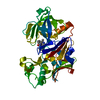
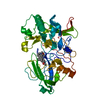
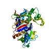
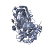
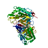
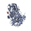
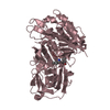
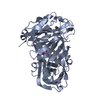
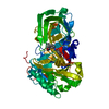
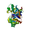
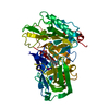
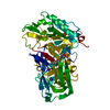
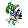
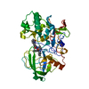
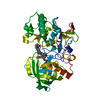
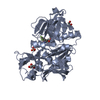
| タイトル | Crystal Structure of Human BACE-1 in Complex with Compound NB-360 (compound 54) |
|---|
 要素 要素 | Beta-secretase 1 |
|---|
 キーワード キーワード | HYDROLASE / Beta-secretase / BACE1 / memaosin2 / aspartic acid proteinase / alzheimer's disease / enzyme-inhibitor complex / Structure-based drug design |
|---|
| 機能・相同性 |  機能・相同性情報 機能・相同性情報
memapsin 2 / Golgi-associated vesicle lumen / beta-aspartyl-peptidase activity / signaling receptor ligand precursor processing / amyloid-beta formation / amyloid precursor protein catabolic process / membrane protein ectodomain proteolysis / amyloid-beta metabolic process / prepulse inhibition / detection of mechanical stimulus involved in sensory perception of pain ...memapsin 2 / Golgi-associated vesicle lumen / beta-aspartyl-peptidase activity / signaling receptor ligand precursor processing / amyloid-beta formation / amyloid precursor protein catabolic process / membrane protein ectodomain proteolysis / amyloid-beta metabolic process / prepulse inhibition / detection of mechanical stimulus involved in sensory perception of pain / response to insulin-like growth factor stimulus / cellular response to manganese ion / multivesicular body / swimming behavior / presynaptic modulation of chemical synaptic transmission / cellular response to copper ion / hippocampal mossy fiber to CA3 synapse / protein serine/threonine kinase binding / trans-Golgi network / protein processing / recycling endosome / response to lead ion / cellular response to amyloid-beta / synaptic vesicle / late endosome / peptidase activity / positive regulation of neuron apoptotic process / amyloid-beta binding / endopeptidase activity / amyloid fibril formation / aspartic-type endopeptidase activity / early endosome / lysosome / endosome membrane / endosome / membrane raft / endoplasmic reticulum lumen / Amyloid fiber formation / axon / neuronal cell body / dendrite / enzyme binding / cell surface / Golgi apparatus / proteolysis / membrane / plasma membrane類似検索 - 分子機能 Beta-secretase BACE1 / Beta-secretase BACE / Memapsin-like / Eukaryotic aspartyl protease / Aspartic peptidase A1 family / Peptidase family A1 domain / Peptidase family A1 domain profile. / Aspartic peptidase, active site / Eukaryotic and viral aspartyl proteases active site. / Aspartic peptidase domain superfamily類似検索 - ドメイン・相同性 |
|---|
| 生物種 |  Homo sapiens (ヒト) Homo sapiens (ヒト) |
|---|
| 手法 |  X線回折 / シンクロトロン / フーリエ合成 / 解像度: 1.94 Å X線回折 / シンクロトロン / フーリエ合成 / 解像度: 1.94 Å |
|---|
 データ登録者 データ登録者 | Rondeau, J.M. / Wirth, E. |
|---|
 引用 引用 | ジャーナル: J.Med.Chem. / 年: 2021
タイトル: Synthesis of the Potent, Selective, and Efficacious beta-Secretase (BACE1) Inhibitor NB-360.
著者: Rueeger, H. / Lueoend, R. / Machauer, R. / Veenstra, S.J. / Holzer, P. / Hurth, K. / Voegtle, M. / Frederiksen, M. / Rondeau, J.M. / Tintelnot-Blomley, M. / Jacobson, L.H. / Staufenbiel, M. / ...著者: Rueeger, H. / Lueoend, R. / Machauer, R. / Veenstra, S.J. / Holzer, P. / Hurth, K. / Voegtle, M. / Frederiksen, M. / Rondeau, J.M. / Tintelnot-Blomley, M. / Jacobson, L.H. / Staufenbiel, M. / Laue, G. / Neumann, U. |
|---|
| 履歴 | | 登録 | 2020年11月25日 | 登録サイト: PDBE / 処理サイト: PDBE |
|---|
| 改定 1.0 | 2021年4月28日 | Provider: repository / タイプ: Initial release |
|---|
| 改定 1.1 | 2021年5月5日 | Group: Database references / カテゴリ: citation / citation_author
Item: _citation.journal_volume / _citation.page_first ..._citation.journal_volume / _citation.page_first / _citation.page_last / _citation_author.identifier_ORCID |
|---|
| 改定 1.2 | 2024年10月23日 | Group: Data collection / Database references / Structure summary
カテゴリ: chem_comp_atom / chem_comp_bond ...chem_comp_atom / chem_comp_bond / database_2 / pdbx_entry_details / pdbx_modification_feature
Item: _database_2.pdbx_DOI / _database_2.pdbx_database_accession / _pdbx_entry_details.has_protein_modification |
|---|
|
|---|
 ムービー
ムービー コントローラー


 データを開く
 基本情報
基本情報 構造の表示
構造の表示 ダウンロードとリンク
 その他のダウンロード
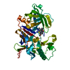


 PDBj
PDBj

 集合体
集合体

 分子量: 449.401 Da / 分子数: 1 / 由来タイプ: 合成 / 式: C21H19F4N5O2 / タイプ: SUBJECT OF INVESTIGATION
 分子量: 18.015 Da / 分子数: 245 / 由来タイプ: 天然 / 式: H2O
 試料調製
試料調製 / ビームライン: X10SA / 波長: 1 Å
 解析
解析