National Institutes of Health/National Institute of General Medical Sciences (NIH/NIGMS)
R01 GM30598
米国
National Institutes of Health/National Heart, Lung, and Blood Institute (NIH/NHLBI)
P01 HL110869
米国
National Institutes of Health/National Institute of Arthritis and Musculoskeletal and Skin Diseases (NIH/NIAMS)
R01 AR53975
米国
American Heart Association
15PRE25090150
米国
National Institutes of Health/National Center for Research Resources (NIH/NCRR)
S10 RR25080
米国
National Institutes of Health/Office of the Director
S10 OD018142
米国
引用
ジャーナル: J Struct Biol / 年: 2017 タイトル: The structure of the actin-smooth muscle myosin motor domain complex in the rigor state. 著者: Chaity Banerjee / Zhongjun Hu / Zhong Huang / J Anthony Warrington / Dianne W Taylor / Kathleen M Trybus / Susan Lowey / Kenneth A Taylor / 要旨: Myosin-based motility utilizes catalysis of ATP to drive the relative sliding of F-actin and myosin. The earliest detailed model based on cryo-electron microscopy (cryoEM) and X-ray crystallography ...Myosin-based motility utilizes catalysis of ATP to drive the relative sliding of F-actin and myosin. The earliest detailed model based on cryo-electron microscopy (cryoEM) and X-ray crystallography postulated that higher actin affinity and lever arm movement were coupled to closure of a feature of the myosin head dubbed the actin-binding cleft. Several studies since then using crystallography of myosin-V and cryoEM structures of F-actin bound myosin-I, -II and -V have provided details of this model. The smooth muscle myosin II interaction with F-actin may differ from those for striated and non-muscle myosin II due in part to different lengths of important surface loops. Here we report a ∼6 Å resolution reconstruction of F-actin decorated with the nucleotide-free recombinant smooth muscle myosin-II motor domain (MD) from images recorded using a direct electron detector. Resolution is highest for F-actin and the actin-myosin interface (3.5-4 Å) and lowest (∼6-7 Å) for those parts of the MD at the highest radius. Atomic models built into the F-actin density are quite comparable to those previously reported for rabbit muscle actin and show density from the bound ADP. The atomic model of the MD, is quite similar to a recently published structure of vertebrate non-muscle myosin II bound to F-actin and a crystal structure of nucleotide free myosin-V. Larger differences are observed when compared to the cryoEM structure of F-actin decorated with rabbit skeletal muscle myosin subfragment 1. The differences suggest less closure of the 50 kDa domain in the actin bound skeletal muscle myosin structure.
全体 : Filamentous muscle alpha-actin decorated with the motor domain of...
全体
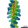
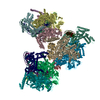
名称: Filamentous muscle alpha-actin decorated with the motor domain of recombinant smooth muscle myosin II motor domain expressed in Sf9 cells
要素
複合体: Filamentous muscle alpha-actin decorated with the motor domain of recombinant smooth muscle myosin II motor domain expressed in Sf9 cells
複合体: alpha-actin
タンパク質・ペプチド: Myosin-11
複合体: myosin II
タンパク質・ペプチド: Actin, alpha skeletal muscle
リガンド: ADENOSINE-5'-DIPHOSPHATE
リガンド: MAGNESIUM ION
-
超分子 #1: Filamentous muscle alpha-actin decorated with the motor domain of...
超分子
名称: Filamentous muscle alpha-actin decorated with the motor domain of recombinant smooth muscle myosin II motor domain expressed in Sf9 cells タイプ: complex / ID: 1 / 親要素: 0 / 含まれる分子: #1-#2
pH: 7.4 詳細: actin buffer: 10 mM imidazole, 10 mM KCl, 1.0 mM MgCl2, 1.0 mM EGTA, 0.5 mM DTT, pH 7.4, myosin buffer: 10 mM imidazole, 10 mM KCl, 1.0 mM MgCl2, 1.0 mM EGTA, 0.5 mM DTT, pH 7.0
凍結剤: ETHANE / チャンバー内湿度: 100 % / チャンバー内温度: 276 K / 装置: GATAN CRYOPLUNGE 3 詳細: Some specimens were frozen manually using a homemade plunger..
-
電子顕微鏡法
顕微鏡
FEI TITAN KRIOS
撮影
フィルム・検出器のモデル: DIRECT ELECTRON DE-20 (5k x 3k) 検出モード: INTEGRATING / デジタル化 - 画像ごとのフレーム数: 1-43 / 実像数: 4000 / 平均露光時間: 1.34 sec. / 平均電子線量: 60.0 e/Å2 / 詳細: Only 1417 of the 4000 recorded images were used.
選択した数: 346395 詳細: Each "particle" consisted of a filament segment masked to a length of 21.0 nm, or slightly more than 7 actin subunits with 2.76 nm separation. Adjacent filament segments overlapped by about 6 ...詳細: Each "particle" consisted of a filament segment masked to a length of 21.0 nm, or slightly more than 7 actin subunits with 2.76 nm separation. Adjacent filament segments overlapped by about 6 subunit repeats (approximately 84% overlap).
 ムービー
ムービー コントローラー


 データを開く
 基本情報
基本情報 マップデータ
 試料
試料 キーワード
キーワード 機能・相同性情報

 データ登録者
 米国, 6件
  引用
引用 構造の表示
構造の表示
 ダウンロードとリンク
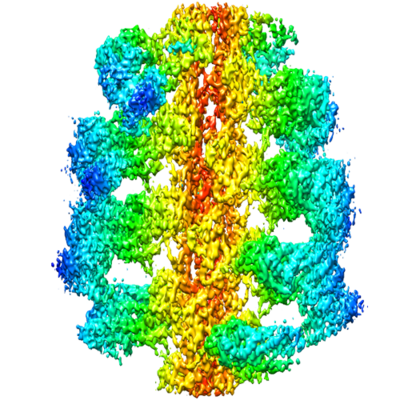 emd_7100.png
 http://ftp.pdbj.org/pub/emdb/structures/EMD-7100


 Z (Sec.)
 Y (Row.)
 X (Col.)


 試料の構成要素
 Spodoptera frugiperda (ツマジロクサヨトウ)
 解析
解析 電子顕微鏡法
 FIELD EMISSION GUN