 Movie
Movie Controller

[English] 日本語
 Yorodumi
- PDB-6bih: The Structure of the Actin-Smooth Muscle Myosin Motor Domain Comp... -
+ Open data

- Basic information
| Entry | Database: PDB / ID: 6bih | |||||||||||||||||||||
|---|---|---|---|---|---|---|---|---|---|---|---|---|---|---|---|---|---|---|---|---|---|---|
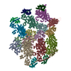
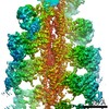
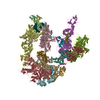
| Title | The Structure of the Actin-Smooth Muscle Myosin Motor Domain Complex in the Rigor State | |||||||||||||||||||||
 Components Components |
| |||||||||||||||||||||
 Keywords Keywords | MOTOR PROTEIN / ADP-F-actin / apo-myosin / helix muscle | |||||||||||||||||||||
| Function / homology |  Function and homology information Function and homology informationelastic fiber assembly / skeletal muscle myosin thick filament assembly / myofibril assembly / myosin light chain binding / myosin II binding / muscle myosin complex / myosin filament / actomyosin structure organization / myosin II complex / cardiac muscle cell development ...elastic fiber assembly / skeletal muscle myosin thick filament assembly / myofibril assembly / myosin light chain binding / myosin II binding / muscle myosin complex / myosin filament / actomyosin structure organization / myosin II complex / cardiac muscle cell development / structural constituent of muscle / cytoskeletal motor activator activity / microfilament motor activity / myosin heavy chain binding / tropomyosin binding / actin filament bundle / troponin I binding / filamentous actin / mesenchyme migration / myofibril / skeletal muscle myofibril / smooth muscle contraction / actin filament bundle assembly / striated muscle thin filament / skeletal muscle thin filament assembly / actin monomer binding / skeletal muscle fiber development / stress fiber / titin binding / actin filament polymerization / filopodium / actin filament / ADP binding / Hydrolases; Acting on acid anhydrides; Acting on acid anhydrides to facilitate cellular and subcellular movement / calcium-dependent protein binding / actin filament binding / lamellipodium / actin binding / cell body / calmodulin binding / protein domain specific binding / hydrolase activity / calcium ion binding / positive regulation of gene expression / magnesium ion binding / ATP binding / identical protein binding / cytoplasm Similarity search - Function | |||||||||||||||||||||
| Biological species |   | |||||||||||||||||||||
| Method | ELECTRON MICROSCOPY / helical reconstruction / cryo EM / Resolution: 6 Å | |||||||||||||||||||||
 Authors Authors | Taylor, K.A. / Banerjee, C. / Hu, Z. | |||||||||||||||||||||
| Funding support |  United States, 6items United States, 6items
| |||||||||||||||||||||
 Citation Citation | Journal: J Struct Biol / Year: 2017 Title: The structure of the actin-smooth muscle myosin motor domain complex in the rigor state. Authors: Chaity Banerjee / Zhongjun Hu / Zhong Huang / J Anthony Warrington / Dianne W Taylor / Kathleen M Trybus / Susan Lowey / Kenneth A Taylor / Abstract: Myosin-based motility utilizes catalysis of ATP to drive the relative sliding of F-actin and myosin. The earliest detailed model based on cryo-electron microscopy (cryoEM) and X-ray crystallography ...Myosin-based motility utilizes catalysis of ATP to drive the relative sliding of F-actin and myosin. The earliest detailed model based on cryo-electron microscopy (cryoEM) and X-ray crystallography postulated that higher actin affinity and lever arm movement were coupled to closure of a feature of the myosin head dubbed the actin-binding cleft. Several studies since then using crystallography of myosin-V and cryoEM structures of F-actin bound myosin-I, -II and -V have provided details of this model. The smooth muscle myosin II interaction with F-actin may differ from those for striated and non-muscle myosin II due in part to different lengths of important surface loops. Here we report a ∼6 Å resolution reconstruction of F-actin decorated with the nucleotide-free recombinant smooth muscle myosin-II motor domain (MD) from images recorded using a direct electron detector. Resolution is highest for F-actin and the actin-myosin interface (3.5-4 Å) and lowest (∼6-7 Å) for those parts of the MD at the highest radius. Atomic models built into the F-actin density are quite comparable to those previously reported for rabbit muscle actin and show density from the bound ADP. The atomic model of the MD, is quite similar to a recently published structure of vertebrate non-muscle myosin II bound to F-actin and a crystal structure of nucleotide free myosin-V. Larger differences are observed when compared to the cryoEM structure of F-actin decorated with rabbit skeletal muscle myosin subfragment 1. The differences suggest less closure of the 50 kDa domain in the actin bound skeletal muscle myosin structure. | |||||||||||||||||||||
| History |
|
- Structure visualization
| Movie |
Movie viewer |
|---|---|
| Structure viewer | Molecule: MolmilJmol/JSmol |
- Downloads & links
-Download
| PDBx/mmCIF format | 6bih.cif.gz | 373.9 KB | Display | PDBx/mmCIF format |
|---|---|---|---|---|
| PDB format | pdb6bih.ent.gz | 297.9 KB | Display | PDB format |
| PDBx/mmJSON format | 6bih.json.gz | Tree view | PDBx/mmJSON format | |
| Others |  Other downloads Other downloads |
-Validation report
| Arichive directory | https://data.pdbj.org/pub/pdb/validation_reports/bi/6bihftp://data.pdbj.org/pub/pdb/validation_reports/bi/6bih | HTTPS FTP |
|---|
-Related structure data
| Related structure data |  7100MC M: map data used to model this data C: citing same article ( |
|---|---|
| Similar structure data |


-Links
 PDBj
PDBj


- Assembly
| Deposited unit | 
|
|---|---|
| 1 | x 7
|
| 2 |
|
| Symmetry | Helical symmetry: (Circular symmetry: 1 / Dyad axis: no / N subunits divisor: 1 / Num. of operations: 7 / Rise per n subunits: 28.18685 Å / Rotation per n subunits: -166.77931 °) |
-Components
| #1: Protein | Mass: 91343.227 Da / Num. of mol.: 1 Source method: isolated from a genetically manipulated source Source: (gene. exp.)   Spodoptera frugiperda (fall armyworm) / References: UniProt: P10587 Spodoptera frugiperda (fall armyworm) / References: UniProt: P10587 |
|---|---|
| #2: Protein | Mass: 42096.953 Da / Num. of mol.: 1 / Source method: isolated from a natural source / Source: (natural) |
| #3: Chemical | ChemComp-ADP /   Mass: 427.201 Da / Num. of mol.: 1 / Source method: obtained synthetically / Formula: C10H15N5O10P2 / Comment: ADP, energy-carrying molecule*YM Mass: 427.201 Da / Num. of mol.: 1 / Source method: obtained synthetically / Formula: C10H15N5O10P2 / Comment: ADP, energy-carrying molecule*YM |
| #4: Chemical | ChemComp-MG /   Mass: 24.305 Da / Num. of mol.: 1 / Source method: obtained synthetically / Formula: Mg Mass: 24.305 Da / Num. of mol.: 1 / Source method: obtained synthetically / Formula: Mg |
-Experimental details
-Experiment
| Experiment | Method: ELECTRON MICROSCOPY |
|---|---|
| EM experiment | Aggregation state: FILAMENT / 3D reconstruction method: helical reconstruction |
- Sample preparation
| Component |
| ||||||||||||||||||||||||
|---|---|---|---|---|---|---|---|---|---|---|---|---|---|---|---|---|---|---|---|---|---|---|---|---|---|
| Molecular weight | Experimental value: NO | ||||||||||||||||||||||||
| Source (natural) |
| ||||||||||||||||||||||||
| Source (recombinant) | Organism: Spodoptera frugiperda (fall armyworm) | ||||||||||||||||||||||||
| Buffer solution | pH: 7.4 Details: actin buffer: 10 mM imidazole, 10 mM KCl, 1.0 mM MgCl2, 1.0 mM EGTA, 0.5 mM DTT, pH 7.4, myosin buffer: 10 mM imidazole, 10 mM KCl, 1.0 mM MgCl2, 1.0 mM EGTA, 0.5 mM DTT, pH 7.0 | ||||||||||||||||||||||||
| Specimen | Conc.: 0.1 mg/ml / Embedding applied: NO / Shadowing applied: NO / Staining applied: NO / Vitrification applied: YES | ||||||||||||||||||||||||
| Specimen support | Grid material: COPPER / Grid mesh size: 200 divisions/in. / Grid type: Quantifoil R2/1 | ||||||||||||||||||||||||
| Vitrification | Instrument: GATAN CRYOPLUNGE 3 / Cryogen name: ETHANE / Humidity: 100 % / Chamber temperature: 276 K Details: Some specimens were frozen manually using a homemade plunger. |
- Electron microscopy imaging
| Experimental equipment |  Model: Titan Krios / Image courtesy: FEI Company |
|---|---|
| Microscopy | Model: FEI TITAN KRIOS |
| Electron gun | Electron source:  FIELD EMISSION GUN / Accelerating voltage: 300 kV / Illumination mode: FLOOD BEAM FIELD EMISSION GUN / Accelerating voltage: 300 kV / Illumination mode: FLOOD BEAM |
| Electron lens | Mode: BRIGHT FIELD / Nominal defocus max: 5000 nm / Nominal defocus min: 2200 nm / Alignment procedure: COMA FREE |
| Specimen holder | Cryogen: NITROGEN / Specimen holder model: FEI TITAN KRIOS AUTOGRID HOLDER |
| Image recording | Average exposure time: 1.34 sec. / Electron dose: 60 e/Å2 / Detector mode: INTEGRATING / Film or detector model: DIRECT ELECTRON DE-20 (5k x 3k) / Num. of real images: 4000 / Details: Only 1417 of the 4000 recorded images were used. |
| Image scans | Movie frames/image: 43 / Used frames/image: 1-43 |
- Processing
| EM software |
| ||||||||||||||||||||||||||||||||||||
|---|---|---|---|---|---|---|---|---|---|---|---|---|---|---|---|---|---|---|---|---|---|---|---|---|---|---|---|---|---|---|---|---|---|---|---|---|---|
| CTF correction | Type: PHASE FLIPPING ONLY | ||||||||||||||||||||||||||||||||||||
| Helical symmerty | Angular rotation/subunit: -166.77931 ° / Axial rise/subunit: 28.18685 Å / Axial symmetry: C1 | ||||||||||||||||||||||||||||||||||||
| Particle selection | Num. of particles selected: 346395 Details: Each "particle" consisted of a filament segment masked to a length of 21.0 nm, or slightly more than 7 actin subunits with 2.76 nm separation. Adjacent filament segments overlapped by about ...Details: Each "particle" consisted of a filament segment masked to a length of 21.0 nm, or slightly more than 7 actin subunits with 2.76 nm separation. Adjacent filament segments overlapped by about 6 subunit repeats (approximately 84% overlap). | ||||||||||||||||||||||||||||||||||||
| 3D reconstruction | Resolution: 6 Å / Resolution method: FSC 0.143 CUT-OFF / Num. of particles: 85000 / Algorithm: EXACT BACK PROJECTION / Num. of class averages: 4 / Symmetry type: HELICAL | ||||||||||||||||||||||||||||||||||||
| Atomic model building | B value: 390.86 / Protocol: FLEXIBLE FIT / Space: REAL |
