 Movie
Movie Controller

[English] 日本語
 Yorodumi
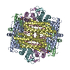
- PDB-6zgl: Structure of DPS determined by movement-free cryoEM with zero dos... -
+ Open data

- Basic information
| Entry | Database: PDB / ID: 6zgl | ||||||
|---|---|---|---|---|---|---|---|
| Title | Structure of DPS determined by movement-free cryoEM with zero dose extrapolation | ||||||
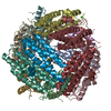
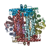
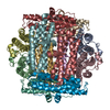
 Components Components | DNA protection during starvation protein | ||||||
 Keywords Keywords | DNA BINDING PROTEIN / DNA-BINDING PROTEIN | ||||||
| Function / homology |  Function and homology information Function and homology informationDnaA-Dps complex / Oxidoreductases; Oxidizing metal ions / oxidoreductase activity, acting on metal ions / nucleoid / chromosome condensation / response to starvation / response to stress / negative regulation of DNA-templated DNA replication initiation / ferric iron binding / intracellular iron ion homeostasis ...DnaA-Dps complex / Oxidoreductases; Oxidizing metal ions / oxidoreductase activity, acting on metal ions / nucleoid / chromosome condensation / response to starvation / response to stress / negative regulation of DNA-templated DNA replication initiation / ferric iron binding / intracellular iron ion homeostasis / DNA binding / identical protein binding / membrane / cytoplasm Similarity search - Function | ||||||
| Biological species |  | ||||||
| Method | ELECTRON MICROSCOPY / single particle reconstruction / cryo EM / Resolution: 1.9 Å | ||||||
 Authors Authors | Naydenova, K. / Russo, C.J. | ||||||
| Funding support |  United Kingdom, 1items United Kingdom, 1items
| ||||||
 Citation Citation | Journal: Science / Year: 2020 Title: Cryo-EM with sub-1 Å specimen movement. Authors: Katerina Naydenova / Peipei Jia / Christopher J Russo /  Abstract: Most information loss in cryogenic electron microscopy (cryo-EM) stems from particle movement during imaging, which remains poorly understood. We show that this movement is caused by buckling and ...Most information loss in cryogenic electron microscopy (cryo-EM) stems from particle movement during imaging, which remains poorly understood. We show that this movement is caused by buckling and subsequent deformation of the suspended ice, with a threshold that depends directly on the shape of the frozen water layer set by the support foil. We describe a specimen support design that eliminates buckling and reduces electron beam-induced particle movement to less than 1 angstrom. The design allows precise foil tracking during imaging with high-speed detectors, thereby lessening demands on cryostage precision and stability. It includes a maximal density of holes, which increases throughput in automated cryo-EM without degrading data quality. Movement-free imaging allows extrapolation to a three-dimensional map of the specimen at zero electron exposure, before the onset of radiation damage. | ||||||
| History |
|
- Structure visualization
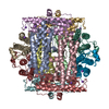
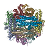
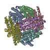
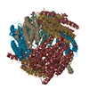
| Movie |
Movie viewer |
|---|---|
| Structure viewer | Molecule: MolmilJmol/JSmol |
- Downloads & links
-Download
| PDBx/mmCIF format | 6zgl.cif.gz | 380.4 KB | Display | PDBx/mmCIF format |
|---|---|---|---|---|
| PDB format | pdb6zgl.ent.gz | 311.1 KB | Display | PDB format |
| PDBx/mmJSON format | 6zgl.json.gz | Tree view | PDBx/mmJSON format | |
| Others |  Other downloads Other downloads |
-Validation report
| Summary document | 6zgl_validation.pdf.gz | 886.9 KB | Display | wwPDB validaton report |
|---|---|---|---|---|
| Full document | 6zgl_full_validation.pdf.gz | 909 KB | Display | |
| Data in XML | 6zgl_validation.xml.gz | 49.6 KB | Display | |
| Data in CIF | 6zgl_validation.cif.gz | 71.2 KB | Display | |
| Arichive directory | https://data.pdbj.org/pub/pdb/validation_reports/zg/6zglftp://data.pdbj.org/pub/pdb/validation_reports/zg/6zgl | HTTPS FTP |
-Related structure data
| Related structure data | 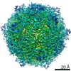 11210MC M: map data used to model this data C: citing same article ( |
|---|---|
| Similar structure data | |
| EM raw data | EMPIAR-10445 (Title: Movies of DPS in 260 nm gold foil holes, which eliminate specimen movement Data size: 1.5 TB Data #1: Unaligned multi-frame micrographs of DPS in 260 nm hole supports [micrographs - multiframe]) |

-Links
 PDBj
PDBj


- Assembly
| Deposited unit | 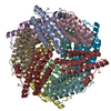
|
|---|---|
| 1 |
|
-Components
| #1: Protein | Mass: 18720.295 Da / Num. of mol.: 12 Source method: isolated from a genetically manipulated source Source: (gene. exp.) #2: Water | ChemComp-HOH / |  Mass: 18.015 Da / Num. of mol.: 964 / Source method: isolated from a natural source / Formula: H2O Mass: 18.015 Da / Num. of mol.: 964 / Source method: isolated from a natural source / Formula: H2O |
|---|
-Experimental details
-Experiment
| Experiment | Method: ELECTRON MICROSCOPY |
|---|---|
| EM experiment | Aggregation state: PARTICLE / 3D reconstruction method: single particle reconstruction |
- Sample preparation
| Component | Name: DNA protection during starvation protein (DPS) / Type: COMPLEX / Entity ID: #1 / Source: RECOMBINANT |
|---|---|
| Molecular weight | Experimental value: NO |
| Source (natural) | Organism: |
| Source (recombinant) | Organism: |
| Buffer solution | pH: 7.7 |
| Specimen | Embedding applied: NO / Shadowing applied: NO / Staining applied: NO / Vitrification applied: YES |
| Vitrification | Cryogen name: ETHANE |
- Electron microscopy imaging
| Experimental equipment |  Model: Titan Krios / Image courtesy: FEI Company |
|---|---|
| Microscopy | Model: FEI TITAN KRIOS |
| Electron gun | Electron source:  FIELD EMISSION GUN / Accelerating voltage: 300 kV / Illumination mode: FLOOD BEAM FIELD EMISSION GUN / Accelerating voltage: 300 kV / Illumination mode: FLOOD BEAM |
| Electron lens | Mode: BRIGHT FIELD |
| Image recording | Electron dose: 35 e/Å2 / Film or detector model: FEI FALCON IV (4k x 4k) |
- Processing
| Software | Name: REFMAC / Version: 5.8.0256 / Classification: refinement | ||||||||||||||||||||||||||||||||||||||||||||||||||||||||||||||||||||||||||||||||||||||||||||||||||||||||||
|---|---|---|---|---|---|---|---|---|---|---|---|---|---|---|---|---|---|---|---|---|---|---|---|---|---|---|---|---|---|---|---|---|---|---|---|---|---|---|---|---|---|---|---|---|---|---|---|---|---|---|---|---|---|---|---|---|---|---|---|---|---|---|---|---|---|---|---|---|---|---|---|---|---|---|---|---|---|---|---|---|---|---|---|---|---|---|---|---|---|---|---|---|---|---|---|---|---|---|---|---|---|---|---|---|---|---|---|
| CTF correction | Type: PHASE FLIPPING AND AMPLITUDE CORRECTION | ||||||||||||||||||||||||||||||||||||||||||||||||||||||||||||||||||||||||||||||||||||||||||||||||||||||||||
| Symmetry | Point symmetry: T (tetrahedral) | ||||||||||||||||||||||||||||||||||||||||||||||||||||||||||||||||||||||||||||||||||||||||||||||||||||||||||
| 3D reconstruction | Resolution: 1.9 Å / Resolution method: FSC 0.143 CUT-OFF / Num. of particles: 275623 / Symmetry type: POINT | ||||||||||||||||||||||||||||||||||||||||||||||||||||||||||||||||||||||||||||||||||||||||||||||||||||||||||
| Refinement | Resolution: 1.9→1.9 Å / Cor.coef. Fo:Fc: 0.764 / ESU R: 0.168 Stereochemistry target values: MAXIMUM LIKELIHOOD WITH PHASES Details: HYDROGENS HAVE BEEN ADDED IN THE RIDING POSITIONS
| ||||||||||||||||||||||||||||||||||||||||||||||||||||||||||||||||||||||||||||||||||||||||||||||||||||||||||
| Solvent computation | Ion probe radii: 0.8 Å / Shrinkage radii: 0.8 Å / VDW probe radii: 1.2 Å / Solvent model: MASK | ||||||||||||||||||||||||||||||||||||||||||||||||||||||||||||||||||||||||||||||||||||||||||||||||||||||||||
| Displacement parameters | Biso mean: 19.636 Å2
| ||||||||||||||||||||||||||||||||||||||||||||||||||||||||||||||||||||||||||||||||||||||||||||||||||||||||||
| Refinement step | Cycle: 1 / Total: 15616 | ||||||||||||||||||||||||||||||||||||||||||||||||||||||||||||||||||||||||||||||||||||||||||||||||||||||||||
| Refine LS restraints |
|