 Movie
Movie Controller

[English] 日本語
 Yorodumi
- PDB-6ybt: Structure of a human 48S translational initiation complex - eIF3bgi -
+ Open data

- Basic information
| Entry | Database: PDB / ID: 6ybt | ||||||||||||
|---|---|---|---|---|---|---|---|---|---|---|---|---|---|
| Title | Structure of a human 48S translational initiation complex - eIF3bgi | ||||||||||||
 Components Components |
| ||||||||||||
 Keywords Keywords | TRANSLATION / eIF3 / ribosome / initiation complex | ||||||||||||
| Function / homology |  Function and homology information Function and homology informationviral translational termination-reinitiation / eukaryotic translation initiation factor 3 complex, eIF3e / eukaryotic translation initiation factor 3 complex, eIF3m / IRES-dependent viral translational initiation / translation reinitiation / formation of cytoplasmic translation initiation complex / multi-eIF complex / eukaryotic translation initiation factor 3 complex / eukaryotic 43S preinitiation complex / eukaryotic 48S preinitiation complex ...viral translational termination-reinitiation / eukaryotic translation initiation factor 3 complex, eIF3e / eukaryotic translation initiation factor 3 complex, eIF3m / IRES-dependent viral translational initiation / translation reinitiation / formation of cytoplasmic translation initiation complex / multi-eIF complex / eukaryotic translation initiation factor 3 complex / eukaryotic 43S preinitiation complex / eukaryotic 48S preinitiation complex / regulation of translational initiation / cytoplasmic translational initiation / Formation of the ternary complex, and subsequently, the 43S complex / Ribosomal scanning and start codon recognition / Translation initiation complex formation / Formation of a pool of free 40S subunits / GTP hydrolysis and joining of the 60S ribosomal subunit / L13a-mediated translational silencing of Ceruloplasmin expression / translation initiation factor activity / translation initiation factor binding / translational initiation / negative regulation of ERK1 and ERK2 cascade / receptor tyrosine kinase binding / cytoplasmic stress granule / molecular adaptor activity / microtubule / postsynaptic density / mRNA binding / synapse / nucleolus / structural molecule activity / RNA binding / extracellular exosome / nucleoplasm / membrane / cytoplasm / cytosol Similarity search - Function | ||||||||||||
| Biological species |  Homo sapiens (human) Homo sapiens (human) | ||||||||||||
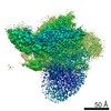
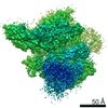
| Method | ELECTRON MICROSCOPY / single particle reconstruction / cryo EM / Resolution: 6 Å | ||||||||||||
 Authors Authors | Brito Querido, J. / Sokabe, M. / Kraatz, S. / Gordiyenko, Y. / Skehel, M. / Fraser, C. / Ramakrishnan, V. | ||||||||||||
| Funding support |  United Kingdom, United Kingdom,  United States, 3items United States, 3items
| ||||||||||||
 Citation Citation | Journal: Science / Year: 2020 Title: Structure of a human 48 translational initiation complex. Authors: Jailson Brito Querido / Masaaki Sokabe / Sebastian Kraatz / Yuliya Gordiyenko / J Mark Skehel / Christopher S Fraser / V Ramakrishnan / Abstract: A key step in translational initiation is the recruitment of the 43 preinitiation complex by the cap-binding complex [eukaryotic initiation factor 4F (eIF4F)] at the 5' end of messenger RNA (mRNA) to ...A key step in translational initiation is the recruitment of the 43 preinitiation complex by the cap-binding complex [eukaryotic initiation factor 4F (eIF4F)] at the 5' end of messenger RNA (mRNA) to form the 48 initiation complex (i.e., the 48). The 48 then scans along the mRNA to locate a start codon. To understand the mechanisms involved, we used cryo-electron microscopy to determine the structure of a reconstituted human 48 The structure reveals insights into early events of translation initiation complex assembly, as well as how eIF4F interacts with subunits of eIF3 near the mRNA exit channel in the 43 The location of eIF4F is consistent with a slotting model of mRNA recruitment and suggests that downstream mRNA is unwound at least in part by being "pulled" through the 40 subunit during scanning. | ||||||||||||
| History |
|
- Structure visualization
| Movie |
Movie viewer |
|---|---|
| Structure viewer | Molecule: MolmilJmol/JSmol |
- Downloads & links
-Download
| PDBx/mmCIF format | 6ybt.cif.gz | 196 KB | Display | PDBx/mmCIF format |
|---|---|---|---|---|
| PDB format | pdb6ybt.ent.gz | 115.7 KB | Display | PDB format |
| PDBx/mmJSON format | 6ybt.json.gz | Tree view | PDBx/mmJSON format | |
| Others |  Other downloads Other downloads |
-Validation report
| Arichive directory | https://data.pdbj.org/pub/pdb/validation_reports/yb/6ybtftp://data.pdbj.org/pub/pdb/validation_reports/yb/6ybt | HTTPS FTP |
|---|
-Related structure data
| Related structure data |  10773MC  6ybdC 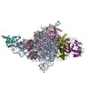 6ybsC  6ybvC 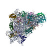 6ybwC  6zmwC M: map data used to model this data C: citing same article ( |
|---|---|
| Similar structure data |


-Links
 PDBj
PDBj


- Assembly
| Deposited unit | 
|
|---|---|
| 1 |
|
-Components
| #1: Protein | Mass: 163179.531 Da / Num. of mol.: 1 / Source method: isolated from a natural source / Source: (natural) Homo sapiens (human) / References: UniProt: Q14152 |
|---|---|
| #2: Protein | Mass: 36543.773 Da / Num. of mol.: 1 / Source method: isolated from a natural source / Source: (natural) Homo sapiens (human) / References: UniProt: Q13347 |
| #3: Protein | Mass: 92593.414 Da / Num. of mol.: 1 / Source method: isolated from a natural source / Source: (natural) Homo sapiens (human) / References: UniProt: P55884 |
-Experimental details
-Experiment
| Experiment | Method: ELECTRON MICROSCOPY |
|---|---|
| EM experiment | Aggregation state: PARTICLE / 3D reconstruction method: single particle reconstruction |
- Sample preparation
| Component | Name: Eukaryotic translation initiation factor / Type: RIBOSOME / Entity ID: all / Source: NATURAL |
|---|---|
| Source (natural) | Organism: Homo sapiens (human) |
| Buffer solution | pH: 7.4 |
| Specimen | Embedding applied: NO / Shadowing applied: NO / Staining applied: NO / Vitrification applied: YES |
| Vitrification | Instrument: FEI VITROBOT MARK IV / Cryogen name: ETHANE / Humidity: 100 % |
- Electron microscopy imaging
| Experimental equipment |  Model: Titan Krios / Image courtesy: FEI Company |
|---|---|
| Microscopy | Model: FEI TITAN KRIOS |
| Electron gun | Electron source:  FIELD EMISSION GUN / Accelerating voltage: 300 kV / Illumination mode: FLOOD BEAM FIELD EMISSION GUN / Accelerating voltage: 300 kV / Illumination mode: FLOOD BEAM |
| Electron lens | Mode: BRIGHT FIELD |
| Image recording | Average exposure time: 1 sec. / Electron dose: 107 e/Å2 / Detector mode: INTEGRATING / Film or detector model: FEI FALCON III (4k x 4k) |
- Processing
| Software |
| ||||||||||||||||||||||||
|---|---|---|---|---|---|---|---|---|---|---|---|---|---|---|---|---|---|---|---|---|---|---|---|---|---|
| CTF correction | Type: PHASE FLIPPING AND AMPLITUDE CORRECTION | ||||||||||||||||||||||||
| 3D reconstruction | Resolution: 6 Å / Resolution method: FSC 0.143 CUT-OFF / Num. of particles: 144882 / Symmetry type: POINT | ||||||||||||||||||||||||
| Refinement | Cross valid method: NONE Stereochemistry target values: GeoStd + Monomer Library + CDL v1.2 | ||||||||||||||||||||||||
| Displacement parameters | Biso mean: 616.69 Å2 | ||||||||||||||||||||||||
| Refine LS restraints |
|
