 Movie
Movie Controller


+ Open data

- Basic information
| Entry | Database: PDB / ID: 6y3y | |||||||||
|---|---|---|---|---|---|---|---|---|---|---|
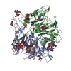
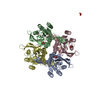
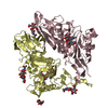
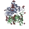
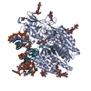
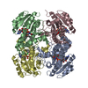
| Title | Human Coronavirus HKU1 Haemagglutinin-Esterase | |||||||||
 Components Components | Hemagglutinin-esterase | |||||||||
 Keywords Keywords | VIRAL PROTEIN / Coronavirus / Glycoprotein / Nidovirus / Esterase | |||||||||
| Function / homology |  Function and homology information Function and homology informationsialate 9-O-acetylesterase activity / sialate 4-O-acetylesterase activity / sialate O-acetylesterase activity / sialate O-acetylesterase / host cell surface receptor binding / fusion of virus membrane with host plasma membrane / viral envelope / host cell plasma membrane / virion membrane / membrane Similarity search - Function | |||||||||
| Biological species |  Human coronavirus HKU1 Human coronavirus HKU1 | |||||||||
| Method | ELECTRON MICROSCOPY / single particle reconstruction / cryo EM / Resolution: 3.39 Å | |||||||||
 Authors Authors | Hurdiss, D.L. / Drulyte, I. / Pronker, M.F. | |||||||||
 Citation Citation | Journal: Nat Commun / Year: 2020 Title: Cryo-EM structure of coronavirus-HKU1 haemagglutinin esterase reveals architectural changes arising from prolonged circulation in humans. Authors: Daniel L Hurdiss / Ieva Drulyte / Yifei Lang / Tatiana M Shamorkina / Matti F Pronker / Frank J M van Kuppeveld / Joost Snijder / Raoul J de Groot /  Abstract: The human betacoronaviruses HKU1 and OC43 (subgenus Embecovirus) arose from separate zoonotic introductions, OC43 relatively recently and HKU1 apparently much longer ago. Embecovirus particles ...The human betacoronaviruses HKU1 and OC43 (subgenus Embecovirus) arose from separate zoonotic introductions, OC43 relatively recently and HKU1 apparently much longer ago. Embecovirus particles contain two surface projections called spike (S) and haemagglutinin-esterase (HE), with S mediating receptor binding and membrane fusion, and HE acting as a receptor-destroying enzyme. Together, they promote dynamic virion attachment to glycan-based receptors, specifically 9-O-acetylated sialic acid. Here we present the cryo-EM structure of the ~80 kDa, heavily glycosylated HKU1 HE at 3.4 Å resolution. Comparison with existing HE structures reveals a drastically truncated lectin domain, incompatible with sialic acid binding, but with the structure and function of the esterase domain left intact. Cryo-EM and mass spectrometry analysis reveals a putative glycan shield on the now redundant lectin domain. The findings further our insight into the evolution and host adaptation of human embecoviruses, and demonstrate the utility of cryo-EM for studying small, heavily glycosylated proteins. #1: Journal: Biorxiv / Year: 2020Title: Cryo-EM structure of coronavirus-HKU1 haemagglutinin esterase reveals architectural changes arising from prolonged circulation in humans Authors: Hurdiss, D.L. / Drulyte, I. / Lang, Y. / Shamorkina, T.M. / Pronker, M.F. / van Kuppeveld, F.J.M. / Snijder, J. / de Groot, R.J. | |||||||||
| History |
|
- Structure visualization
| Movie |
Movie viewer |
|---|---|
| Structure viewer | Molecule: MolmilJmol/JSmol |
- Downloads & links
-Download
| PDBx/mmCIF format | 6y3y.cif.gz | 144.9 KB | Display | PDBx/mmCIF format |
|---|---|---|---|---|
| PDB format | pdb6y3y.ent.gz | 110.6 KB | Display | PDB format |
| PDBx/mmJSON format | 6y3y.json.gz | Tree view | PDBx/mmJSON format | |
| Others |  Other downloads Other downloads |
-Validation report
| Arichive directory | https://data.pdbj.org/pub/pdb/validation_reports/y3/6y3yftp://data.pdbj.org/pub/pdb/validation_reports/y3/6y3y | HTTPS FTP |
|---|
-Related structure data
| Related structure data | 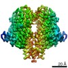 10676MC M: map data used to model this data C: citing same article ( |
|---|---|
| Similar structure data | |
| EM raw data | EMPIAR-10390 (Title: Cryo-EM structure of coronavirus-HKU1 haemagglutinin esterase Data size: 5.0 TB Data #1: Unaligned multi-frame gain-normalised movies of HCoV-HKU1 HE [micrographs - multiframe]) |


-Links
 PDBj
PDBj


- Assembly
| Deposited unit | 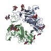
|
|---|---|
| 1 |
|
-Components
-Protein , 1 types, 2 molecules AB
| #1: Protein | Mass: 40432.648 Da / Num. of mol.: 2 Source method: isolated from a genetically manipulated source Source: (gene. exp.) Human coronavirus HKU1 / Gene: HE, 2 / Cell line (production host): HEK293T / Production host:  Homo sapiens (human) / References: UniProt: Q5MQD1, sialate O-acetylesterase Homo sapiens (human) / References: UniProt: Q5MQD1, sialate O-acetylesterase |
|---|
-Sugars , 5 types, 14 molecules 
| #2: Polysaccharide | Source method: isolated from a genetically manipulated source #3: Polysaccharide | Source method: isolated from a genetically manipulated source #4: Polysaccharide | Source method: isolated from a genetically manipulated source #5: Polysaccharide | Source method: isolated from a genetically manipulated source #6: Sugar | ChemComp-NAG /  Type: D-saccharide, beta linking / Mass: 221.208 Da / Num. of mol.: 6 Type: D-saccharide, beta linking / Mass: 221.208 Da / Num. of mol.: 6Source method: isolated from a genetically manipulated source Formula: C8H15NO6 |
|---|
-Details
| Has ligand of interest | N |
|---|---|
| Has protein modification | Y |
-Experimental details
-Experiment
| Experiment | Method: ELECTRON MICROSCOPY |
|---|---|
| EM experiment | Aggregation state: PARTICLE / 3D reconstruction method: single particle reconstruction |
- Sample preparation
| Component | Name: Human Coronavirus HKU1 Haemagglutinin-Esterase / Type: COMPLEX / Details: Dimeric complex / Entity ID: #1 / Source: RECOMBINANT | |||||||||||||||
|---|---|---|---|---|---|---|---|---|---|---|---|---|---|---|---|---|
| Molecular weight |
| |||||||||||||||
| Source (natural) | Organism: Human coronavirus HKU1 | |||||||||||||||
| Source (recombinant) | Organism: Homo sapiens (human) | |||||||||||||||
| Buffer solution | pH: 8 | |||||||||||||||
| Buffer component |
| |||||||||||||||
| Specimen | Conc.: 4 mg/ml / Embedding applied: NO / Shadowing applied: NO / Staining applied: NO / Vitrification applied: YES | |||||||||||||||
| Specimen support | Details: 20 mA / Grid material: COPPER / Grid mesh size: 200 divisions/in. / Grid type: Quantifoil R1.2/1.3 | |||||||||||||||
| Vitrification | Instrument: FEI VITROBOT MARK IV / Cryogen name: ETHANE / Humidity: 95 % / Chamber temperature: 277 K / Details: Blot force = 1 Blot time = 5.5 seconds |
- Electron microscopy imaging
| Experimental equipment |  Model: Titan Krios / Image courtesy: FEI Company |
|---|---|
| Microscopy | Model: TFS KRIOS Details: Titan Krios G4 was used with fringe-free imaging and aberration-free image shift. |
| Electron gun | Electron source:  FIELD EMISSION GUN / Accelerating voltage: 300 kV / Illumination mode: FLOOD BEAM FIELD EMISSION GUN / Accelerating voltage: 300 kV / Illumination mode: FLOOD BEAM |
| Electron lens | Mode: BRIGHT FIELD / Nominal magnification: 96000 X / Nominal defocus max: -2500 nm / Nominal defocus min: -1000 nm / Cs: 2.7 mm / C2 aperture diameter: 50 µm / Alignment procedure: COMA FREE |
| Specimen holder | Cryogen: NITROGEN / Specimen holder model: FEI TITAN KRIOS AUTOGRID HOLDER |
| Image recording | Average exposure time: 5.6 sec. / Electron dose: 40 e/Å2 / Film or detector model: OTHER / Num. of grids imaged: 1 / Num. of real images: 6029 / Details: Falcon 4 Direct Electron Detector |
| Image scans | Sampling size: 14 µm / Width: 4096 / Height: 4096 |
- Processing
| Software |
| ||||||||||||||||||||||||||||||||||||||||
|---|---|---|---|---|---|---|---|---|---|---|---|---|---|---|---|---|---|---|---|---|---|---|---|---|---|---|---|---|---|---|---|---|---|---|---|---|---|---|---|---|---|
| EM software |
| ||||||||||||||||||||||||||||||||||||||||
| Image processing | Details: Falcon 4 Direct Electron Detector | ||||||||||||||||||||||||||||||||||||||||
| CTF correction | Type: PHASE FLIPPING AND AMPLITUDE CORRECTION | ||||||||||||||||||||||||||||||||||||||||
| Particle selection | Num. of particles selected: 936260 / Details: Relion autopicking | ||||||||||||||||||||||||||||||||||||||||
| Symmetry | Point symmetry: C2 (2 fold cyclic) | ||||||||||||||||||||||||||||||||||||||||
| 3D reconstruction | Resolution: 3.39 Å / Resolution method: FSC 0.143 CUT-OFF / Num. of particles: 119717 / Algorithm: BACK PROJECTION / Symmetry type: POINT | ||||||||||||||||||||||||||||||||||||||||
| Atomic model building | Protocol: RIGID BODY FIT / Space: REAL / Target criteria: Correlation coefficient Details: A homology model of HCOV-HKU1 HE (uniprot ID: Q5MQD1) was generated using the phyre2 server | ||||||||||||||||||||||||||||||||||||||||
| Refinement | Stereochemistry target values: GeoStd + Monomer Library | ||||||||||||||||||||||||||||||||||||||||
| Refine LS restraints |
|
