[English] 日本語
 Yorodumi
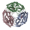
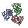
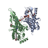
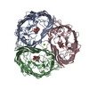
- PDB-6xwq: Structure of glutamate transporter homologue GltTk in the saturat... -
+ Open data
- Basic information
| Entry | Database: PDB / ID: 6xwq | ||||||||||||||||||||||||||||||
|---|---|---|---|---|---|---|---|---|---|---|---|---|---|---|---|---|---|---|---|---|---|---|---|---|---|---|---|---|---|---|---|
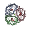
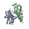
| Title | Structure of glutamate transporter homologue GltTk in the saturated conditions | ||||||||||||||||||||||||||||||
 Components Components | Proton/glutamate symporter, SDF family | ||||||||||||||||||||||||||||||
 Keywords Keywords | TRANSPORT PROTEIN / AMINO ACID TRANSPORTER / ASPARTATE TRANSPORT / GLUTAMATE TRANSPORTER HOMOLOGUE / MEMBRANE PROTEIN | ||||||||||||||||||||||||||||||
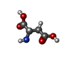
| Function / homology |  Function and homology information Function and homology informationcarboxylic acid transport / symporter activity / membrane / plasma membrane Similarity search - Function | ||||||||||||||||||||||||||||||
| Biological species |   Thermococcus kodakarensis (archaea) Thermococcus kodakarensis (archaea) | ||||||||||||||||||||||||||||||
| Method | ELECTRON MICROSCOPY / single particle reconstruction / cryo EM / Resolution: 3.41 Å | ||||||||||||||||||||||||||||||
 Authors Authors | Arkhipova, V. / Slotboom, D.J. / Guskov, A. | ||||||||||||||||||||||||||||||
 Citation Citation |  Journal: Nat Commun / Year: 2020 Journal: Nat Commun / Year: 2020Title: Structural ensemble of a glutamate transporter homologue in lipid nanodisc environment. Authors: Valentina Arkhipova / Albert Guskov / Dirk J Slotboom /   Abstract: Glutamate transporters are cation-coupled secondary active membrane transporters that clear the neurotransmitter L-glutamate from the synaptic cleft. These transporters are homotrimers, with each ...Glutamate transporters are cation-coupled secondary active membrane transporters that clear the neurotransmitter L-glutamate from the synaptic cleft. These transporters are homotrimers, with each protomer functioning independently by an elevator-type mechanism, in which a mobile transport domain alternates between inward- and outward-oriented states. Using single-particle cryo-EM we have determined five structures of the glutamate transporter homologue Glt, a Na- L-aspartate symporter, embedded in lipid nanodiscs. Dependent on the substrate concentrations used, the protomers of the trimer adopt a variety of asymmetrical conformations, consistent with the independent movement. Six of the 15 resolved protomers are in a hitherto elusive state of the transport cycle in which the inward-facing transporters are loaded with Na ions. These structures explain how substrate-leakage is prevented - a strict requirement for coupled transport. The belt protein of the lipid nanodiscs bends around the inward oriented protomers, suggesting that membrane deformations occur during transport. | ||||||||||||||||||||||||||||||
| History |
|
- Structure visualization
| Movie |
 Movie viewer Movie viewer |
|---|---|
| Structure viewer | Molecule:  Molmil Molmil Jmol/JSmol Jmol/JSmol |
- Downloads & links
- Download
| PDBx/mmCIF format |  6xwq.cif.gz 6xwq.cif.gz | 212.7 KB | Display |  PDBx/mmCIF format PDBx/mmCIF format |
|---|---|---|---|---|
| PDB format |  pdb6xwq.ent.gz pdb6xwq.ent.gz | 172.9 KB | Display |  PDB format PDB format |
| PDBx/mmJSON format |  6xwq.json.gz 6xwq.json.gz | Tree view |  PDBx/mmJSON format PDBx/mmJSON format | |
| Others |  Other downloads Other downloads |
-Validation report
| Summary document |  6xwq_validation.pdf.gz 6xwq_validation.pdf.gz | 857.1 KB | Display |  wwPDB validaton report wwPDB validaton report |
|---|---|---|---|---|
| Full document |  6xwq_full_validation.pdf.gz 6xwq_full_validation.pdf.gz | 875.6 KB | Display | |
| Data in XML |  6xwq_validation.xml.gz 6xwq_validation.xml.gz | 39.1 KB | Display | |
| Data in CIF |  6xwq_validation.cif.gz 6xwq_validation.cif.gz | 59 KB | Display | |
| Arichive directory |  https://data.pdbj.org/pub/pdb/validation_reports/xw/6xwq https://data.pdbj.org/pub/pdb/validation_reports/xw/6xwq ftp://data.pdbj.org/pub/pdb/validation_reports/xw/6xwq ftp://data.pdbj.org/pub/pdb/validation_reports/xw/6xwq | HTTPS FTP |
-Related structure data
| Related structure data |  10635MC  6xwnC  6xwoC  6xwpC  6xwrC M: map data used to model this data C: citing same article ( |
|---|---|
| Similar structure data |
- Links
Links
- Assembly
| Deposited unit | 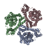
|
|---|---|
| 1 |
|
- Components
| #1: Protein | Mass: 45580.203 Da / Num. of mol.: 3 Source method: isolated from a genetically manipulated source Source: (gene. exp.)   Thermococcus kodakarensis (strain ATCC BAA-918 / JCM 12380 / KOD1) (archaea) Thermococcus kodakarensis (strain ATCC BAA-918 / JCM 12380 / KOD1) (archaea)Gene: TK0986 / Production host:  #2: Chemical | Has ligand of interest | N | Has protein modification | N | |
|---|
-Experimental details
-Experiment
| Experiment | Method: ELECTRON MICROSCOPY |
|---|---|
| EM experiment | Aggregation state: PARTICLE / 3D reconstruction method: single particle reconstruction |
- Sample preparation
| Component | Name: Trimer of GltTk / Type: COMPLEX / Entity ID: #1 / Source: RECOMBINANT |
|---|---|
| Molecular weight | Value: 0.136 MDa / Experimental value: YES |
| Source (natural) | Organism:   Thermococcus kodakarensis KOD1 (archaea) Thermococcus kodakarensis KOD1 (archaea) |
| Source (recombinant) | Organism:  |
| Buffer solution | pH: 7.4 |
| Specimen | Embedding applied: NO / Shadowing applied: NO / Staining applied: NO / Vitrification applied: YES |
| Vitrification | Cryogen name: ETHANE |
- Electron microscopy imaging
| Experimental equipment |  Model: Talos Arctica / Image courtesy: FEI Company |
|---|---|
| Microscopy | Model: FEI TECNAI ARCTICA |
| Electron gun | Electron source:  FIELD EMISSION GUN / Accelerating voltage: 200 kV / Illumination mode: FLOOD BEAM FIELD EMISSION GUN / Accelerating voltage: 200 kV / Illumination mode: FLOOD BEAM |
| Electron lens | Mode: BRIGHT FIELD |
| Image recording | Electron dose: 53 e/Å2 / Detector mode: COUNTING / Film or detector model: GATAN K2 QUANTUM (4k x 4k) |
- Processing
| Software | Name: PHENIX / Version: 1.16_3549: / Classification: refinement | ||||||||||||||||||||||||
|---|---|---|---|---|---|---|---|---|---|---|---|---|---|---|---|---|---|---|---|---|---|---|---|---|---|
| EM software | Name: PHENIX / Category: model refinement | ||||||||||||||||||||||||
| CTF correction | Type: NONE | ||||||||||||||||||||||||
| Symmetry | Point symmetry: C3 (3 fold cyclic) | ||||||||||||||||||||||||
| 3D reconstruction | Resolution: 3.41 Å / Resolution method: FSC 0.143 CUT-OFF / Num. of particles: 65762 / Symmetry type: POINT | ||||||||||||||||||||||||
| Refine LS restraints |
|
 Movie
Movie Controller


 PDBj
PDBj
