 Movie
Movie Controller

[English] 日本語
 Yorodumi
- PDB-6xfm: Molecular structure of the core of amyloid-like fibrils formed by... -
+ Open data

- Basic information
| Entry | Database: PDB / ID: 6xfm | ||||||
|---|---|---|---|---|---|---|---|
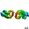
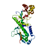
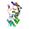
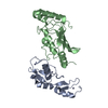
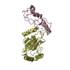
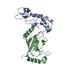
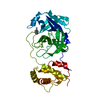
| Title | Molecular structure of the core of amyloid-like fibrils formed by residues 111-214 of FUS | ||||||
 Components Components | RNA-binding protein FUS | ||||||
 Keywords Keywords | RNA BINDING PROTEIN / PROTEIN FIBRIL / Low complexity domain / Protein aggregation / Amyloid Fibril | ||||||
| Function / homology |  Function and homology information Function and homology informationmembraneless organelle assembly / mRNA stabilization / regulation of RNA splicing / Processing of Capped Intron-Containing Pre-mRNA / postsynaptic cytosol / positive regulation of double-strand break repair via homologous recombination / presynaptic cytosol / mRNA Splicing - Major Pathway / RNA splicing / transcription coregulator activity ...membraneless organelle assembly / mRNA stabilization / regulation of RNA splicing / Processing of Capped Intron-Containing Pre-mRNA / postsynaptic cytosol / positive regulation of double-strand break repair via homologous recombination / presynaptic cytosol / mRNA Splicing - Major Pathway / RNA splicing / transcription coregulator activity / mRNA 3'-UTR binding / molecular condensate scaffold activity / protein homooligomerization / GABA-ergic synapse / amyloid fibril formation / transcription coactivator activity / chromatin binding / regulation of transcription by RNA polymerase II / regulation of DNA-templated transcription / glutamatergic synapse / DNA binding / RNA binding / zinc ion binding / nucleoplasm / identical protein binding / nucleus Similarity search - Function | ||||||
| Biological species |  Homo sapiens (human) Homo sapiens (human) | ||||||
| Method | ELECTRON MICROSCOPY / single particle reconstruction / cryo EM / Resolution: 2.62 Å | ||||||
 Authors Authors | Tycko, R. / Lee, M. / Ghosh, U. / Thurber, K. / Kato, M. | ||||||
 Citation Citation | Journal: Nat Commun / Year: 2020 Title: Molecular structure and interactions within amyloid-like fibrils formed by a low-complexity protein sequence from FUS. Authors: Myungwoon Lee / Ujjayini Ghosh / Kent R Thurber / Masato Kato / Robert Tycko /   Abstract: Protein domains without the usual distribution of amino acids, called low complexity (LC) domains, can be prone to self-assembly into amyloid-like fibrils. Self-assembly of LC domains that are nearly ...Protein domains without the usual distribution of amino acids, called low complexity (LC) domains, can be prone to self-assembly into amyloid-like fibrils. Self-assembly of LC domains that are nearly devoid of hydrophobic residues, such as the 214-residue LC domain of the RNA-binding protein FUS, is particularly intriguing from the biophysical perspective and is biomedically relevant due to its occurrence within neurons in amyotrophic lateral sclerosis, frontotemporal dementia, and other neurodegenerative diseases. We report a high-resolution molecular structural model for fibrils formed by the C-terminal half of the FUS LC domain (FUS-LC-C, residues 111-214), based on a density map with 2.62 Å resolution from cryo-electron microscopy (cryo-EM). In the FUS-LC-C fibril core, residues 112-150 adopt U-shaped conformations and form two subunits with in-register, parallel cross-β structures, arranged with quasi-2 symmetry. All-atom molecular dynamics simulations indicate that the FUS-LC-C fibril core is stabilized by a plethora of hydrogen bonds involving sidechains of Gln, Asn, Ser, and Tyr residues, both along and transverse to the fibril growth direction, including diverse sidechain-to-backbone, sidechain-to-sidechain, and sidechain-to-water interactions. Nuclear magnetic resonance measurements additionally show that portions of disordered residues 151-214 remain highly dynamic in FUS-LC-C fibrils and that fibrils formed by the N-terminal half of the FUS LC domain (FUS-LC-N, residues 2-108) have the same core structure as fibrils formed by the full-length LC domain. These results contribute to our understanding of the molecular structural basis for amyloid formation by FUS and by LC domains in general. | ||||||
| History |
|
- Structure visualization
| Movie |
Movie viewer |
|---|---|
| Structure viewer | Molecule: MolmilJmol/JSmol |
- Downloads & links
-Download
| PDBx/mmCIF format | 6xfm.cif.gz | 1.1 MB | Display | PDBx/mmCIF format |
|---|---|---|---|---|
| PDB format | pdb6xfm.ent.gz | 947.5 KB | Display | PDB format |
| PDBx/mmJSON format | 6xfm.json.gz | Tree view | PDBx/mmJSON format | |
| Others |  Other downloads Other downloads |
-Validation report
| Arichive directory | https://data.pdbj.org/pub/pdb/validation_reports/xf/6xfmftp://data.pdbj.org/pub/pdb/validation_reports/xf/6xfm | HTTPS FTP |
|---|
-Related structure data
| Related structure data |  22169MC M: map data used to model this data C: citing same article ( |
|---|---|
| Similar structure data |

-Links
 PDBj
PDBj


- Assembly
| Deposited unit | 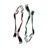
|
|---|---|
| 1 |
|
| Number of models | 14 |
-Components
| #1: Protein | Mass: 10024.784 Da / Num. of mol.: 8 / Fragment: low complexity domain (UNP residues 111-214) Source method: isolated from a genetically manipulated source Source: (gene. exp.) Homo sapiens (human) / Gene: FUS, TLS / Production host:  |
|---|
-Experimental details
-Experiment
| Experiment | Method: ELECTRON MICROSCOPY |
|---|---|
| EM experiment | Aggregation state: HELICAL ARRAY / 3D reconstruction method: single particle reconstruction |
- Sample preparation
| Component | Name: FUS low complexity sequence / Type: COMPLEX Details: C-terminal domain of FUS low complexity domain (111-214) Entity ID: all / Source: RECOMBINANT |
|---|---|
| Molecular weight | Value: 40.4 kDa/nm / Experimental value: YES |
| Source (natural) | Organism: Homo sapiens (human) |
| Source (recombinant) | Organism: |
| Buffer solution | pH: 7.4 / Details: 20 mM 2-mercaptoethanol, 0.1 mM PMSF |
| Buffer component | Conc.: 20 mM / Name: Tris HCl / Formula: C4H11NO3 |
| Specimen | Conc.: 0.4 mg/ml / Embedding applied: NO / Shadowing applied: NO / Staining applied: NO / Vitrification applied: YES |
| Specimen support | Details: The grid was glow discharged immediately before use. Grid material: COPPER / Grid mesh size: 300 divisions/in. / Grid type: Quantifoil R2/2 |
| Vitrification | Instrument: LEICA PLUNGER / Cryogen name: ETHANE / Humidity: 99 % / Chamber temperature: 93 K Details: Preblot for 10 seconds and blot for 5 seconds before plunging |
- Electron microscopy imaging
| Experimental equipment |  Model: Titan Krios / Image courtesy: FEI Company |
|---|---|
| Microscopy | Model: FEI TITAN KRIOS |
| Electron gun | Electron source:  FIELD EMISSION GUN / Accelerating voltage: 300 kV / Illumination mode: FLOOD BEAM FIELD EMISSION GUN / Accelerating voltage: 300 kV / Illumination mode: FLOOD BEAM |
| Electron lens | Mode: BRIGHT FIELD / Nominal magnification: 130000 X / Nominal defocus max: 2500 nm / Nominal defocus min: 1000 nm |
| Specimen holder | Cryogen: NITROGEN |
| Image recording | Average exposure time: 6 sec. / Electron dose: 47 e/Å2 / Detector mode: SUPER-RESOLUTION / Film or detector model: GATAN K2 SUMMIT (4k x 4k) / Num. of grids imaged: 2 / Num. of real images: 2404 Details: 58185 fibril segments were manually selected from 2404 micrographs |
| Image scans | Width: 3800 / Height: 3700 / Movie frames/image: 30 |
- Processing
| EM software |
| ||||||||||||||||||||||||||||||||||||||||||||||||
|---|---|---|---|---|---|---|---|---|---|---|---|---|---|---|---|---|---|---|---|---|---|---|---|---|---|---|---|---|---|---|---|---|---|---|---|---|---|---|---|---|---|---|---|---|---|---|---|---|---|
| Image processing | Details: Gatan Imaging Filter (GIF) Quantum LS | ||||||||||||||||||||||||||||||||||||||||||||||||
| CTF correction | Details: Gctf / Type: NONE | ||||||||||||||||||||||||||||||||||||||||||||||||
| Helical symmerty | Angular rotation/subunit: -2 ° / Axial rise/subunit: 4.8 Å / Axial symmetry: C2 | ||||||||||||||||||||||||||||||||||||||||||||||||
| Particle selection | Num. of particles selected: 499206 Details: 499206 of particles were extracted from the 58185 fibril segments using a 400-pixel box size and 91.6% overlap. | ||||||||||||||||||||||||||||||||||||||||||||||||
| Symmetry | Point symmetry: C1 (asymmetric) | ||||||||||||||||||||||||||||||||||||||||||||||||
| 3D reconstruction | Resolution: 2.62 Å / Resolution method: FSC 0.143 CUT-OFF / Num. of particles: 275520 / Algorithm: FOURIER SPACE Details: 3D refinement and post-processing were performed with 21 (screw) symmetry Num. of class averages: 1 / Symmetry type: POINT | ||||||||||||||||||||||||||||||||||||||||||||||||
| Atomic model building | Protocol: OTHER Details: Manually generated model was fit into the density using PHENIX and UCSF Chimera. Further refinements were performed using Xplor-NIH. |
