 Movie
Movie Controller


+ Open data

- Basic information
| Entry | Database: PDB / ID: 6xfa | |||||||||
|---|---|---|---|---|---|---|---|---|---|---|
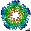
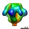
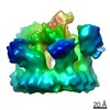
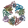
| Title | Cryo-EM structure of EBV BFLF1 | |||||||||
 Components Components | Packaging protein UL32 | |||||||||
 Keywords Keywords | VIRAL PROTEIN / viral packaging / viral cleavage | |||||||||
| Function / homology |  Function and homology information Function and homology informationhost cell cytoplasm / viral envelope / host cell nucleus / zinc ion binding / metal ion binding Similarity search - Function | |||||||||
| Biological species |  Epstein-Barr virus (Epstein-Barr virus) Epstein-Barr virus (Epstein-Barr virus) | |||||||||
| Method | ELECTRON MICROSCOPY / single particle reconstruction / cryo EM / Resolution: 3.6 Å | |||||||||
 Authors Authors | Didychuk, A.L. / Gates, S.N. / Martin, A. / Glaunsinger, B. | |||||||||
| Funding support |  United States, 2items United States, 2items
| |||||||||
 Citation Citation | Journal: Elife / Year: 2021 Title: A pentameric protein ring with novel architecture is required for herpesviral packaging. Authors: Allison L Didychuk / Stephanie N Gates / Matthew R Gardner / Lisa M Strong / Andreas Martin / Britt A Glaunsinger / Abstract: Genome packaging in large double-stranded DNA viruses requires a powerful molecular motor to force the viral genome into nascent capsids, which involves essential accessory factors that are poorly ...Genome packaging in large double-stranded DNA viruses requires a powerful molecular motor to force the viral genome into nascent capsids, which involves essential accessory factors that are poorly understood. Here, we present structures of two such accessory factors from the oncogenic herpesviruses Kaposi's sarcoma-associated herpesvirus (KSHV; ORF68) and Epstein-Barr virus (EBV; BFLF1). These homologous proteins form highly similar homopentameric rings with a positively charged central channel that binds double-stranded DNA. Mutation of individual positively charged residues within but not outside the channel ablates DNA binding, and in the context of KSHV infection, these mutants fail to package the viral genome or produce progeny virions. Thus, we propose a model in which ORF68 facilitates the transfer of newly replicated viral genomes to the packaging motor. | |||||||||
| History |
|
- Structure visualization
| Movie |
Movie viewer |
|---|---|
| Structure viewer | Molecule: MolmilJmol/JSmol |
- Downloads & links
-Download
| PDBx/mmCIF format | 6xfa.cif.gz | 666.8 KB | Display | PDBx/mmCIF format |
|---|---|---|---|---|
| PDB format | pdb6xfa.ent.gz | 552.4 KB | Display | PDB format |
| PDBx/mmJSON format | 6xfa.json.gz | Tree view | PDBx/mmJSON format | |
| Others |  Other downloads Other downloads |
-Validation report
| Arichive directory | https://data.pdbj.org/pub/pdb/validation_reports/xf/6xfaftp://data.pdbj.org/pub/pdb/validation_reports/xf/6xfa | HTTPS FTP |
|---|
-Related structure data
| Related structure data |  22168MC  6xf9C M: map data used to model this data C: citing same article ( |
|---|---|
| Similar structure data |

-Links
 PDBj
PDBj- Assembly
| Deposited unit | 
|
|---|---|
| 1 |
|
-Components
| #1: Protein | Mass: 53678.797 Da / Num. of mol.: 10 Source method: isolated from a genetically manipulated source Source: (gene. exp.) Epstein-Barr virus (strain GD1) (Epstein-Barr virus)Strain: GD1 / Gene: BFLF1 / Production host:  Homo sapiens (human) / References: UniProt: A0A2D1LYN0, UniProt: P03184*PLUS Homo sapiens (human) / References: UniProt: A0A2D1LYN0, UniProt: P03184*PLUS#2: Chemical | ChemComp-ZN /   Mass: 65.409 Da / Num. of mol.: 20 / Source method: obtained synthetically / Formula: Zn Mass: 65.409 Da / Num. of mol.: 20 / Source method: obtained synthetically / Formula: ZnHas ligand of interest | N | |
|---|
-Experimental details
-Experiment
| Experiment | Method: ELECTRON MICROSCOPY |
|---|---|
| EM experiment | Aggregation state: PARTICLE / 3D reconstruction method: single particle reconstruction |
- Sample preparation
| Component | Name: Decamer structure of EBV BFLF1 / Type: COMPLEX / Entity ID: #1 / Source: RECOMBINANT | |||||||||||||||||||||||||||||||||||
|---|---|---|---|---|---|---|---|---|---|---|---|---|---|---|---|---|---|---|---|---|---|---|---|---|---|---|---|---|---|---|---|---|---|---|---|---|
| Molecular weight | Value: 0.536 MDa / Experimental value: NO | |||||||||||||||||||||||||||||||||||
| Source (natural) | Organism: Epstein-Barr virus (strain GD1) (Epstein-Barr virus) | |||||||||||||||||||||||||||||||||||
| Source (recombinant) | Organism: Homo sapiens (human) | |||||||||||||||||||||||||||||||||||
| Buffer solution | pH: 7.6 | |||||||||||||||||||||||||||||||||||
| Buffer component |
| |||||||||||||||||||||||||||||||||||
| Specimen | Conc.: 4 mg/ml / Embedding applied: NO / Shadowing applied: NO / Staining applied: NO / Vitrification applied: YES | |||||||||||||||||||||||||||||||||||
| Specimen support | Grid material: COPPER / Grid mesh size: 300 divisions/in. / Grid type: C-flat | |||||||||||||||||||||||||||||||||||
| Vitrification | Instrument: FEI VITROBOT MARK IV / Cryogen name: ETHANE / Humidity: 90 % / Chamber temperature: 295 K / Details: 2 second blot, 3 second wait |
- Electron microscopy imaging
| Experimental equipment |  Model: Talos Arctica / Image courtesy: FEI Company |
|---|---|
| Microscopy | Model: FEI TALOS ARCTICA |
| Electron gun | Electron source:  FIELD EMISSION GUN / Accelerating voltage: 200 kV / Illumination mode: FLOOD BEAM FIELD EMISSION GUN / Accelerating voltage: 200 kV / Illumination mode: FLOOD BEAM |
| Electron lens | Mode: BRIGHT FIELD / Nominal defocus max: 3000 nm / Nominal defocus min: 1500 nm / Cs: 2.7 mm / C2 aperture diameter: 100 µm |
| Specimen holder | Cryogen: NITROGEN / Specimen holder model: OTHER |
| Image recording | Average exposure time: 2.4 sec. / Electron dose: 48 e/Å2 / Film or detector model: GATAN K3 (6k x 4k) / Num. of grids imaged: 1 / Num. of real images: 839 |
- Processing
| EM software |
| ||||||||||||||||||||||||
|---|---|---|---|---|---|---|---|---|---|---|---|---|---|---|---|---|---|---|---|---|---|---|---|---|---|
| CTF correction | Type: PHASE FLIPPING AND AMPLITUDE CORRECTION | ||||||||||||||||||||||||
| Particle selection | Num. of particles selected: 278234 | ||||||||||||||||||||||||
| Symmetry | Point symmetry: D5 (2x5 fold dihedral) | ||||||||||||||||||||||||
| 3D reconstruction | Resolution: 3.6 Å / Resolution method: FSC 0.143 CUT-OFF / Num. of particles: 32272 / Symmetry type: POINT |
