ムービー
ムービー コントローラー


+ データを開く

- 基本情報
基本情報
| 登録情報 | データベース: PDB / ID: 6ty4 | |||||||||
|---|---|---|---|---|---|---|---|---|---|---|
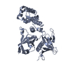
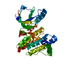
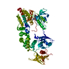
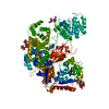
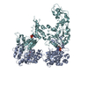
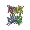
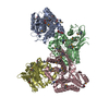
| タイトル | FAK structure with AMP-PNP from single particle analysis of 2D crystals | |||||||||
 要素 要素 | Focal adhesion kinase 1 | |||||||||
 キーワード キーワード | CELL ADHESION / Kinase / Focal Adhesion / Membrane | |||||||||
| 機能・相同性 |  機能・相同性情報 機能・相同性情報Apoptotic cleavage of cellular proteins / NCAM signaling for neurite out-growth / RHO GTPases Activate WASPs and WAVEs / RAF/MAP kinase cascade / Regulation of actin dynamics for phagocytic cup formation / radial glia-guided pyramidal neuron migration / negative regulation of protein autophosphorylation / calcium-dependent cysteine-type endopeptidase activity / positive regulation of substrate-dependent cell migration, cell attachment to substrate / Integrin signaling ...Apoptotic cleavage of cellular proteins / NCAM signaling for neurite out-growth / RHO GTPases Activate WASPs and WAVEs / RAF/MAP kinase cascade / Regulation of actin dynamics for phagocytic cup formation / radial glia-guided pyramidal neuron migration / negative regulation of protein autophosphorylation / calcium-dependent cysteine-type endopeptidase activity / positive regulation of substrate-dependent cell migration, cell attachment to substrate / Integrin signaling / GRB2:SOS provides linkage to MAPK signaling for Integrins / DCC mediated attractive signaling / MET activates PTK2 signaling / Extra-nuclear estrogen signaling / EPHB-mediated forward signaling / p130Cas linkage to MAPK signaling for integrins / VEGFA-VEGFR2 Pathway / angiogenesis involved in wound healing / signal complex assembly / response to pH / negative regulation of cell-substrate adhesion / wound healing, spreading of cells / positive regulation of focal adhesion assembly / negative regulation of anoikis / positive regulation of protein tyrosine kinase activity / regulation of cell adhesion / response to muscle stretch / ciliary basal body / molecular function activator activity / actin filament organization / non-specific protein-tyrosine kinase / non-membrane spanning protein tyrosine kinase activity / epidermal growth factor receptor signaling pathway / sarcolemma / integrin binding / positive regulation of protein binding / cell cortex / protein tyrosine kinase activity / protease binding / protein autophosphorylation / dendritic spine / positive regulation of cell migration / focal adhesion / centrosome / positive regulation of cell population proliferation / perinuclear region of cytoplasm / ATP binding / identical protein binding / nucleus / cytoplasm 類似検索 - 分子機能 | |||||||||
| 生物種 |  | |||||||||
| 手法 | 電子顕微鏡法 / 単粒子再構成法 / クライオ電子顕微鏡法 / 解像度: 5.96 Å | |||||||||
 データ登録者 データ登録者 | Acebron, I. / Righetto, R. / Biyani, N. / Chami, M. / Boskovic, J. / Stahlberg, H. / Lietha, D. | |||||||||
| 資金援助 |  スペイン, スペイン,  スイス, 2件 スイス, 2件
| |||||||||
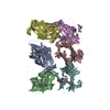
 引用 引用 | ジャーナル: EMBO J / 年: 2020 タイトル: Structural basis of Focal Adhesion Kinase activation on lipid membranes. 著者: Iván Acebrón / Ricardo D Righetto / Christina Schoenherr / Svenja de Buhr / Pilar Redondo / Jayne Culley / Carlos F Rodríguez / Csaba Daday / Nikhil Biyani / Oscar Llorca / Adam Byron / ...著者: Iván Acebrón / Ricardo D Righetto / Christina Schoenherr / Svenja de Buhr / Pilar Redondo / Jayne Culley / Carlos F Rodríguez / Csaba Daday / Nikhil Biyani / Oscar Llorca / Adam Byron / Mohamed Chami / Frauke Gräter / Jasminka Boskovic / Margaret C Frame / Henning Stahlberg / Daniel Lietha /   要旨: Focal adhesion kinase (FAK) is a key component of the membrane proximal signaling layer in focal adhesion complexes, regulating important cellular processes, including cell migration, proliferation, ...Focal adhesion kinase (FAK) is a key component of the membrane proximal signaling layer in focal adhesion complexes, regulating important cellular processes, including cell migration, proliferation, and survival. In the cytosol, FAK adopts an autoinhibited state but is activated upon recruitment into focal adhesions, yet how this occurs or what induces structural changes is unknown. Here, we employ cryo-electron microscopy to reveal how FAK associates with lipid membranes and how membrane interactions unlock FAK autoinhibition to promote activation. Intriguingly, initial binding of FAK to the membrane causes steric clashes that release the kinase domain from autoinhibition, allowing it to undergo a large conformational change and interact itself with the membrane in an orientation that places the active site toward the membrane. In this conformation, the autophosphorylation site is exposed and multiple interfaces align to promote FAK oligomerization on the membrane. We show that interfaces responsible for initial dimerization and membrane attachment are essential for FAK autophosphorylation and resulting cellular activity including cancer cell invasion, while stable FAK oligomerization appears to be needed for optimal cancer cell proliferation in an anchorage-independent manner. Together, our data provide structural details of a key membrane bound state of FAK that is primed for efficient autophosphorylation and activation, hence revealing the critical event in integrin mediated FAK activation and signaling at focal adhesions. | |||||||||
| 履歴 |
|
- 構造の表示
構造の表示
| ムービー |
ムービービューア |
|---|---|
| 構造ビューア | 分子: MolmilJmol/JSmol |
- ダウンロードとリンク
-ダウンロード
| PDBx/mmCIF形式 | 6ty4.cif.gz | 236.8 KB | 表示 | PDBx/mmCIF形式 |
|---|---|---|---|---|
| PDB形式 | pdb6ty4.ent.gz | 189.2 KB | 表示 | PDB形式 |
| PDBx/mmJSON形式 | 6ty4.json.gz | ツリー表示 | PDBx/mmJSON形式 | |
| その他 |  その他のダウンロード その他のダウンロード |
-検証レポート
| 文書・要旨 | 6ty4_validation.pdf.gz | 1.4 MB | 表示 | wwPDB検証レポート |
|---|---|---|---|---|
| 文書・詳細版 | 6ty4_full_validation.pdf.gz | 1.5 MB | 表示 | |
| XML形式データ | 6ty4_validation.xml.gz | 55.8 KB | 表示 | |
| CIF形式データ | 6ty4_validation.cif.gz | 81 KB | 表示 | |
| アーカイブディレクトリ | https://data.pdbj.org/pub/pdb/validation_reports/ty/6ty4ftp://data.pdbj.org/pub/pdb/validation_reports/ty/6ty4 | HTTPS FTP |
-関連構造データ
| 関連構造データ |  10616MC  6ty3C M: このデータのモデリングに利用したマップデータ C: 同じ文献を引用 ( |
|---|---|
| 類似構造データ | |
| 電子顕微鏡画像生データ | EMPIAR-10347 (タイトル: FAK structure from single particle analysis of 2D crystals Data size: 1.8 TB Data #1: Dose-weighted aligned movie averages of the FAK AMP-PNP dataset [micrographs - single frame] Data #2: Unaligned movies of the FAK AMP-PNP dataset [micrographs - multiframe] Data #3: Stack of particles extracted from the FAK AMP-PNP dataset [picked particles - single frame - unprocessed] Data #4: Dose-weighted aligned movie averages of the FAK APO Polara dataset [micrographs - single frame] Data #5: Dose-weighted aligned movie averages of the FAK APO Titan dataset [micrographs - single frame] Data #6: Unaligned movies of the FAK APO Titan dataset [micrographs - multiframe] Data #7: Stack of particles extracted from the FAK APO Titan and Polara datasets [picked particles - single frame - unprocessed]) |


-リンク
 PDBj
PDBj


- 集合体
集合体
| 登録構造単位 | 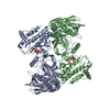
|
|---|---|
| 1 |
|
-要素
| #1: タンパク質 | 分子量: 75476.445 Da / 分子数: 2 / 由来タイプ: 組換発現 / 由来: (組換発現)  Homo sapiens (ヒト) Homo sapiens (ヒト)参照: UniProt: Q00944, non-specific protein-tyrosine kinase #2: 化合物 |   分子量: 24.305 Da / 分子数: 2 / 由来タイプ: 合成 / 式: Mg 分子量: 24.305 Da / 分子数: 2 / 由来タイプ: 合成 / 式: Mg#3: 化合物 |   分子量: 506.196 Da / 分子数: 2 / 由来タイプ: 合成 / 式: C10H17N6O12P3 分子量: 506.196 Da / 分子数: 2 / 由来タイプ: 合成 / 式: C10H17N6O12P3コメント: AMP-PNP, エネルギー貯蔵分子類似体*YM 研究の焦点であるリガンドがあるか | N | |
|---|
-実験情報
-実験
| 実験 | 手法: 電子顕微鏡法 |
|---|---|
| EM実験 | 試料の集合状態: 2D ARRAY / 3次元再構成法: 単粒子再構成法 |
- 試料調製
試料調製
| 構成要素 | 名称: Focal adhesion kinase / タイプ: COMPLEX / Entity ID: #1 / 由来: RECOMBINANT |
|---|---|
| 分子量 | 値: 0.15 MDa / 実験値: NO |
| 由来(天然) | 生物種: |
| 由来(組換発現) | 生物種: Homo sapiens (ヒト) |
| 緩衝液 | pH: 5.5 |
| 試料 | 包埋: NO / シャドウイング: NO / 染色: NO / 凍結: YES |
| 急速凍結 | 凍結剤: ETHANE |
- 電子顕微鏡撮影
| 実験機器 |  モデル: Titan Krios / 画像提供: FEI Company |
|---|---|
| 顕微鏡 | モデル: FEI TITAN KRIOS |
| 電子銃 | 電子線源:  FIELD EMISSION GUN / 加速電圧: 300 kV / 照射モード: FLOOD BEAM FIELD EMISSION GUN / 加速電圧: 300 kV / 照射モード: FLOOD BEAM |
| 電子レンズ | モード: BRIGHT FIELD / Cs: 2.7 mm / C2レンズ絞り径: 100 µm |
| 撮影 | 電子線照射量: 40 e/Å2 / 検出モード: COUNTING フィルム・検出器のモデル: GATAN K2 SUMMIT (4k x 4k) |
- 解析
解析
| EMソフトウェア |
| ||||||||||||||||||||
|---|---|---|---|---|---|---|---|---|---|---|---|---|---|---|---|---|---|---|---|---|---|
| CTF補正 | タイプ: PHASE FLIPPING AND AMPLITUDE CORRECTION | ||||||||||||||||||||
| 3次元再構成 | 解像度: 5.96 Å / 解像度の算出法: FSC 0.143 CUT-OFF / 粒子像の数: 548394 / アルゴリズム: FOURIER SPACE / 対称性のタイプ: POINT | ||||||||||||||||||||
| 原子モデル構築 | プロトコル: FLEXIBLE FIT / 空間: REAL | ||||||||||||||||||||
| 原子モデル構築 | 3D fitting-ID: 1 / PDB chain-ID: A / Source name: PDB / タイプ: experimental model
|