 Movie
Movie Controller


+ Open data

- Basic information
| Entry | Database: PDB / ID: 6tte | ||||||
|---|---|---|---|---|---|---|---|
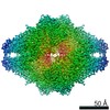
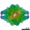
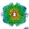
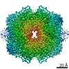
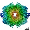
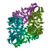
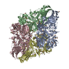
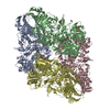
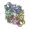
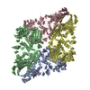
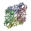
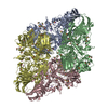
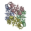
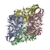
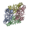
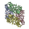
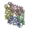
| Title | Beta-galactosidase in complex with PETG | ||||||
 Components Components | Beta-galactosidase | ||||||
 Keywords Keywords | SUGAR BINDING PROTEIN / Bgal / PETG | ||||||
| Function / homology |  Function and homology information Function and homology informationalkali metal ion binding / lactose catabolic process / beta-galactosidase complex / beta-galactosidase / beta-galactosidase activity / carbohydrate binding / magnesium ion binding / identical protein binding Similarity search - Function | ||||||
| Biological species |  | ||||||
| Method | ELECTRON MICROSCOPY / single particle reconstruction / cryo EM / Resolution: 2.2 Å | ||||||
 Authors Authors | Saur, M. / Hartshorn, M.J. / Dong, J. / Reeks, J. / Bunkoczi, G. / Jhoti, H. / Williams, P.A. | ||||||
 Citation Citation | Journal: Drug Discov Today / Year: 2020 Title: Fragment-based drug discovery using cryo-EM. Authors: Michael Saur / Michael J Hartshorn / Jing Dong / Judith Reeks / Gabor Bunkoczi / Harren Jhoti / Pamela A Williams /  Abstract: Recent advances in electron cryo-microscopy (cryo-EM) structure determination have pushed the resolutions obtainable by the method into the range widely considered to be of utility for drug discovery. ...Recent advances in electron cryo-microscopy (cryo-EM) structure determination have pushed the resolutions obtainable by the method into the range widely considered to be of utility for drug discovery. Here, we review the use of cryo-EM in fragment-based drug discovery (FBDD) based on in-house method development. We demonstrate not only that cryo-EM can reveal details of the molecular interactions between fragments and a protein, but also that the current reproducibility, quality, and throughput are compatible with FBDD. We exemplify this using the test system β-galactosidase (Bgal) and the oncology target pyruvate kinase 2 (PKM2). | ||||||
| History |
|
- Structure visualization
| Movie |
Movie viewer |
|---|---|
| Structure viewer | Molecule: MolmilJmol/JSmol |
- Downloads & links
-Download
| PDBx/mmCIF format | 6tte.cif.gz | 832 KB | Display | PDBx/mmCIF format |
|---|---|---|---|---|
| PDB format | pdb6tte.ent.gz | 673.7 KB | Display | PDB format |
| PDBx/mmJSON format | 6tte.json.gz | Tree view | PDBx/mmJSON format | |
| Others |  Other downloads Other downloads |
-Validation report
| Arichive directory | https://data.pdbj.org/pub/pdb/validation_reports/tt/6tteftp://data.pdbj.org/pub/pdb/validation_reports/tt/6tte | HTTPS FTP |
|---|
-Related structure data
| Related structure data | 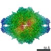 10574MC 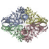 6tshC 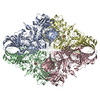 6tskC 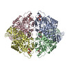 6ttfC  6tthC 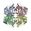 6ttiC 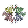 6ttqC C: citing same article ( M: map data used to model this data |
|---|---|
| Similar structure data | |
| EM raw data | EMPIAR-10644 (Title: Beta-galactosidase in complex with PETG / Data size: 720.1 Data #1: Data from EPU (movies have been converted to compressed TIF) [micrographs - multiframe]) |


-Links
 PDBj
PDBj


- Assembly
| Deposited unit | 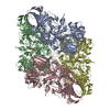
|
|---|---|
| 1 |
|
-Components
| #1: Protein | Mass: 118395.336 Da / Num. of mol.: 4 Source method: isolated from a genetically manipulated source Source: (gene. exp.) References: UniProt: Q8VNN2, UniProt: P00722*PLUS, beta-galactosidase #2: Chemical | ChemComp-MG /   Mass: 24.305 Da / Num. of mol.: 4 / Source method: obtained synthetically / Formula: Mg Mass: 24.305 Da / Num. of mol.: 4 / Source method: obtained synthetically / Formula: Mg#3: Sugar | ChemComp-PTQ /   Type: D-saccharide / Mass: 300.371 Da / Num. of mol.: 4 / Source method: obtained synthetically / Formula: C14H20O5S / Feature type: SUBJECT OF INVESTIGATION Type: D-saccharide / Mass: 300.371 Da / Num. of mol.: 4 / Source method: obtained synthetically / Formula: C14H20O5S / Feature type: SUBJECT OF INVESTIGATION#4: Water | ChemComp-HOH / |  Mass: 18.015 Da / Num. of mol.: 1224 / Source method: isolated from a natural source / Formula: H2O Mass: 18.015 Da / Num. of mol.: 1224 / Source method: isolated from a natural source / Formula: H2OHas ligand of interest | Y | |
|---|
-Experimental details
-Experiment
| Experiment | Method: ELECTRON MICROSCOPY |
|---|---|
| EM experiment | Aggregation state: PARTICLE / 3D reconstruction method: single particle reconstruction |
- Sample preparation
| Component | Name: Beta-galactosidase / Type: COMPLEX / Entity ID: #1 / Source: RECOMBINANT |
|---|---|
| Molecular weight | Value: 0.464 MDa / Experimental value: NO |
| Source (natural) | Organism: |
| Source (recombinant) | Organism: |
| Buffer solution | pH: 6.8 |
| Specimen | Conc.: 0.17 mg/ml / Embedding applied: NO / Shadowing applied: NO / Staining applied: NO / Vitrification applied: YES |
| Specimen support | Grid material: COPPER / Grid mesh size: 300 divisions/in. / Grid type: Quantifoil R1.2/1.3 |
| Vitrification | Instrument: FEI VITROBOT MARK IV / Cryogen name: ETHANE / Humidity: 100 % / Chamber temperature: 277 K |
- Electron microscopy imaging
| Experimental equipment |  Model: Titan Krios / Image courtesy: FEI Company |
|---|---|
| Microscopy | Model: FEI TITAN KRIOS |
| Electron gun | Electron source:  FIELD EMISSION GUN / Accelerating voltage: 300 kV / Illumination mode: FLOOD BEAM FIELD EMISSION GUN / Accelerating voltage: 300 kV / Illumination mode: FLOOD BEAM |
| Electron lens | Mode: BRIGHT FIELD |
| Image recording | Average exposure time: 59.98 sec. / Electron dose: 65.53 e/Å2 / Detector mode: COUNTING / Film or detector model: FEI FALCON III (4k x 4k) / Num. of grids imaged: 1 / Num. of real images: 562 |
- Processing
| EM software | Name: RELION / Version: 3.04 / Category: 3D reconstruction |
|---|---|
| CTF correction | Type: PHASE FLIPPING AND AMPLITUDE CORRECTION |
| Particle selection | Num. of particles selected: 136013 |
| Symmetry | Point symmetry: D2 (2x2 fold dihedral) |
| 3D reconstruction | Resolution: 2.2 Å / Resolution method: FSC 0.143 CUT-OFF / Num. of particles: 49895 / Symmetry type: POINT |
