 Movie
Movie Controller


+ Open data

- Basic information
| Entry | Database: PDB / ID: 6sl0 | ||||||
|---|---|---|---|---|---|---|---|
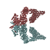
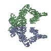
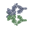
| Title | Complete CtTel1 dimer with C2 symmetry | ||||||
 Components Components | Serine/threonine-protein kinase Tel1 | ||||||
 Keywords Keywords | TRANSFERASE / Kinase / alpha solenoid / PIKK / nucleus / tranferase / dimer / DNA damage signaling | ||||||
| Function / homology |  Function and homology information Function and homology informationchromatin organization / chromosome, telomeric region / non-specific serine/threonine protein kinase / intracellular signal transduction / protein serine kinase activity / DNA repair / protein serine/threonine kinase activity / ATP binding / nucleus Similarity search - Function | ||||||
| Biological species |  Chaetomium thermophilum (fungus) Chaetomium thermophilum (fungus) | ||||||
| Method | ELECTRON MICROSCOPY / single particle reconstruction / cryo EM / Resolution: 3.7 Å | ||||||
 Authors Authors | Jansma, M. / Eustermann, S.E. / Kostrewa, D. / Lammens, K. / Hopfner, K.P. | ||||||
| Funding support |  Germany, 1items Germany, 1items
| ||||||
 Citation Citation | Journal: Structure / Year: 2020 Title: Near-Complete Structure and Model of Tel1ATM from Chaetomium thermophilum Reveals a Robust Autoinhibited ATP State. Authors: Marijke Jansma / Christian Linke-Winnebeck / Sebastian Eustermann / Katja Lammens / Dirk Kostrewa / Kristina Stakyte / Claudia Litz / Brigitte Kessler / Karl-Peter Hopfner / Abstract: Tel1 (ATM in humans) is a large kinase that resides in the cell in an autoinhibited dimeric state and upon activation orchestrates the cellular response to DNA damage. We report the structure of an ...Tel1 (ATM in humans) is a large kinase that resides in the cell in an autoinhibited dimeric state and upon activation orchestrates the cellular response to DNA damage. We report the structure of an endogenous Tel1 dimer from Chaetomium thermophilum. Major parts are at 2.8 Å resolution, including the kinase active site with ATPγS bound, and two different N-terminal solenoid conformations are at 3.4 Å and 3.6 Å, providing a side-chain model for 90% of the Tel1 polypeptide. We show that the N-terminal solenoid has DNA binding activity, but that its movements are not coupled to kinase activation. Although ATPγS and catalytic residues are poised for catalysis, the kinase resides in an autoinhibited state. The PIKK regulatory domain acts as a pseudo-substrate, blocking direct access to the site of catalysis. The structure allows mapping of human cancer mutations and defines mechanisms of autoinhibition at near-atomic resolution. | ||||||
| History |
|
- Structure visualization
| Movie |
Movie viewer |
|---|---|
| Structure viewer | Molecule: MolmilJmol/JSmol |
- Downloads & links
-Download
| PDBx/mmCIF format | 6sl0.cif.gz | 897.8 KB | Display | PDBx/mmCIF format |
|---|---|---|---|---|
| PDB format | pdb6sl0.ent.gz | 702.5 KB | Display | PDB format |
| PDBx/mmJSON format | 6sl0.json.gz | Tree view | PDBx/mmJSON format | |
| Others |  Other downloads Other downloads |
-Validation report
| Arichive directory | https://data.pdbj.org/pub/pdb/validation_reports/sl/6sl0ftp://data.pdbj.org/pub/pdb/validation_reports/sl/6sl0 | HTTPS FTP |
|---|
-Related structure data
| Related structure data | 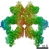 10233MC  6skyC  6skzC  6sl1C M: map data used to model this data C: citing same article ( |
|---|---|
| Similar structure data |


-Links
 PDBj
PDBj

- Assembly
| Deposited unit | 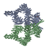
|
|---|---|
| 1 |
|
-Components
| #1: Protein | Mass: 329927.031 Da / Num. of mol.: 2 / Source method: isolated from a natural source Source: (natural) Chaetomium thermophilum (strain DSM 1495 / CBS 144.50 / IMI 039719) (fungus)Strain: DSM 1495 / CBS 144.50 / IMI 039719 References: UniProt: G0S4S9, non-specific serine/threonine protein kinase #2: Chemical |   Mass: 523.247 Da / Num. of mol.: 2 / Source method: obtained synthetically / Formula: C10H16N5O12P3S / Comment: ATP-gamma-S, energy-carrying molecule analogue*YM Mass: 523.247 Da / Num. of mol.: 2 / Source method: obtained synthetically / Formula: C10H16N5O12P3S / Comment: ATP-gamma-S, energy-carrying molecule analogue*YM#3: Chemical |   Mass: 24.305 Da / Num. of mol.: 2 / Source method: obtained synthetically / Formula: Mg Mass: 24.305 Da / Num. of mol.: 2 / Source method: obtained synthetically / Formula: MgHas ligand of interest | N | Has protein modification | Y | |
|---|
-Experimental details
-Experiment
| Experiment | Method: ELECTRON MICROSCOPY |
|---|---|
| EM experiment | Aggregation state: PARTICLE / 3D reconstruction method: single particle reconstruction |
- Sample preparation
| Component | Name: CtTel1 / Type: COMPLEX / Entity ID: #1 / Source: NATURAL | ||||||||||||
|---|---|---|---|---|---|---|---|---|---|---|---|---|---|
| Molecular weight | Value: 0.64 MDa / Experimental value: YES | ||||||||||||
| Source (natural) | Organism: Chaetomium thermophilum var. thermophilum DSM 1495 (fungus) | ||||||||||||
| Buffer solution | pH: 7.5 Details: 1 mM MgCl2 and 0.1 mM ATPgS (final concentrations) added before plunging | ||||||||||||
| Buffer component |
| ||||||||||||
| Specimen | Conc.: 0.15 mg/ml / Embedding applied: NO / Shadowing applied: NO / Staining applied: NO / Vitrification applied: YES | ||||||||||||
| Vitrification | Instrument: LEICA EM GP / Cryogen name: ETHANE / Humidity: 95 % / Chamber temperature: 288.15 K Details: TWEEN-20 was added to a final concentration of 0.05% immediately before vitrification. Sample was preincubated 45 seconds before plunging into liquid ethane. |
- Electron microscopy imaging
| Experimental equipment |  Model: Titan Krios / Image courtesy: FEI Company |
|---|---|
| Microscopy | Model: FEI TITAN KRIOS |
| Electron gun | Electron source:  FIELD EMISSION GUN / Accelerating voltage: 300 kV / Illumination mode: FLOOD BEAM FIELD EMISSION GUN / Accelerating voltage: 300 kV / Illumination mode: FLOOD BEAM |
| Electron lens | Mode: BRIGHT FIELD |
| Image recording | Average exposure time: 8 sec. / Electron dose: 55.5 e/Å2 / Detector mode: COUNTING / Film or detector model: GATAN K2 SUMMIT (4k x 4k) / Num. of grids imaged: 2 / Num. of real images: 13786 |
| Image scans | Movie frames/image: 40 |
- Processing
| Software | Name: PHENIX / Version: 1.16_3549: / Classification: refinement | |||||||||||||||||||||||||
|---|---|---|---|---|---|---|---|---|---|---|---|---|---|---|---|---|---|---|---|---|---|---|---|---|---|---|
| EM software |
| |||||||||||||||||||||||||
| CTF correction | Type: PHASE FLIPPING AND AMPLITUDE CORRECTION | |||||||||||||||||||||||||
| Particle selection | Num. of particles selected: 863937 | |||||||||||||||||||||||||
| Symmetry | Point symmetry: C2 (2 fold cyclic) | |||||||||||||||||||||||||
| 3D reconstruction | Resolution: 3.7 Å / Resolution method: FSC 0.143 CUT-OFF / Num. of particles: 32764 / Symmetry type: POINT | |||||||||||||||||||||||||
| Refinement | Cross valid method: NONE |
