 ムービー
ムービー コントローラー


+ データを開く

- 基本情報
基本情報
| 登録情報 | データベース: PDB / ID: 6r4r | |||||||||||||||
|---|---|---|---|---|---|---|---|---|---|---|---|---|---|---|---|---|
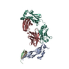
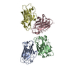
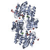
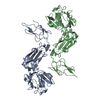
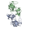
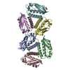
| タイトル | Cryo-EM Structure of the PI3-Kinase SH3 Domain Amyloid Fibril | |||||||||||||||
 要素 要素 | Phosphatidylinositol 3-kinase regulatory subunit alpha | |||||||||||||||
 キーワード キーワード | PROTEIN FIBRIL / amyloid fibril / sh3 domain | |||||||||||||||
| 機能・相同性 |  機能・相同性情報 機能・相同性情報RHOC GTPase cycle / PI3K events in ERBB4 signaling / Interleukin-7 signaling / GAB1 signalosome / PI3K events in ERBB2 signaling / MET activates PI3K/AKT signaling / CDC42 GTPase cycle / RHOD GTPase cycle / RHOJ GTPase cycle / RAC3 GTPase cycle ...RHOC GTPase cycle / PI3K events in ERBB4 signaling / Interleukin-7 signaling / GAB1 signalosome / PI3K events in ERBB2 signaling / MET activates PI3K/AKT signaling / CDC42 GTPase cycle / RHOD GTPase cycle / RHOJ GTPase cycle / RAC3 GTPase cycle / Erythropoietin activates Phosphoinositide-3-kinase (PI3K) / FLT3 Signaling / RND2 GTPase cycle / RND1 GTPase cycle / IRS-mediated signalling / GPVI-mediated activation cascade / Signaling by SCF-KIT / Downstream signal transduction / PI3K/AKT activation / Signaling by ALK / Role of phospholipids in phagocytosis / Tie2 Signaling / Role of LAT2/NTAL/LAB on calcium mobilization / Costimulation by the CD28 family / CD28 dependent PI3K/Akt signaling / RAC1 GTPase cycle / RAC2 GTPase cycle / Interleukin receptor SHC signaling / RND3 GTPase cycle / PI-3K cascade:FGFR1 / PI-3K cascade:FGFR2 / PI-3K cascade:FGFR3 / PI-3K cascade:FGFR4 / Antigen activates B Cell Receptor (BCR) leading to generation of second messengers / PI3K Cascade / PIP3 activates AKT signaling / GP1b-IX-V activation signalling / RAF/MAP kinase cascade / PI5P, PP2A and IER3 Regulate PI3K/AKT Signaling / Synthesis of PIPs at the plasma membrane / RHOA GTPase cycle / RHOF GTPase cycle / DAP12 signaling / RHOU GTPase cycle / RHOV GTPase cycle / Regulation of signaling by CBL / Downstream TCR signaling / RHOG GTPase cycle / RET signaling / Interleukin-3, Interleukin-5 and GM-CSF signaling / VEGFA-VEGFR2 Pathway / phosphatidylinositol 3-kinase regulator activity / phosphatidylinositol 3-kinase activator activity / 1-phosphatidylinositol-3-kinase regulator activity / positive regulation of endoplasmic reticulum unfolded protein response / ErbB-3 class receptor binding / transmembrane receptor protein tyrosine kinase adaptor activity / phosphatidylinositol 3-kinase complex, class IA / phosphatidylinositol 3-kinase complex / enzyme-substrate adaptor activity / Extra-nuclear estrogen signaling / G alpha (q) signalling events / intracellular glucose homeostasis / phosphatidylinositol phosphate biosynthetic process / insulin receptor substrate binding / phosphatidylinositol 3-kinase binding / insulin-like growth factor receptor binding / response to endoplasmic reticulum stress / phosphatidylinositol 3-kinase/protein kinase B signal transduction / substrate adhesion-dependent cell spreading / positive regulation of RNA splicing / insulin-like growth factor receptor signaling pathway / positive regulation of D-glucose import / positive regulation of protein localization to plasma membrane / insulin receptor binding / cellular response to insulin stimulus / positive regulation of protein import into nucleus / protein transport / insulin receptor signaling pathway / protein stabilization / negative regulation of apoptotic process / positive regulation of transcription by RNA polymerase II / identical protein binding / nucleus 類似検索 - 分子機能 | |||||||||||||||
| 生物種 |  | |||||||||||||||
| 手法 | 電子顕微鏡法 / らせん対称体再構成法 / クライオ電子顕微鏡法 / 解像度: 3.4 Å | |||||||||||||||
 データ登録者 データ登録者 | Roeder, C. / Vettore, N. / Mangels, L.N. / Gremer, L. / Ravelli, R.B.G. / Willbold, D. / Hoyer, W. / Buell, A.K. / Schroder, G.F. | |||||||||||||||
| 資金援助 |  ドイツ, 4件 ドイツ, 4件
| |||||||||||||||
 引用 引用 | ジャーナル: Nat Commun / 年: 2019 タイトル: Atomic structure of PI3-kinase SH3 amyloid fibrils by cryo-electron microscopy. 著者: Christine Röder / Nicola Vettore / Lena N Mangels / Lothar Gremer / Raimond B G Ravelli / Dieter Willbold / Wolfgang Hoyer / Alexander K Buell / Gunnar F Schröder /   要旨: High resolution structural information on amyloid fibrils is crucial for the understanding of their formation mechanisms and for the rational design of amyloid inhibitors in the context of protein ...High resolution structural information on amyloid fibrils is crucial for the understanding of their formation mechanisms and for the rational design of amyloid inhibitors in the context of protein misfolding diseases. The Src-homology 3 domain of phosphatidyl-inositol-3-kinase (PI3K-SH3) is a model amyloid system that plays a pivotal role in our basic understanding of protein misfolding and aggregation. Here, we present the atomic model of the PI3K-SH3 amyloid fibril with a resolution determined to 3.4 Å by cryo-electron microscopy (cryo-EM). The fibril is composed of two intertwined protofilaments that create an interface spanning 13 residues from each monomer. The model comprises residues 1-77 out of 86 amino acids in total, with the missing residues located in the highly flexible C-terminus. The fibril structure allows us to rationalise the effects of chemically conservative point mutations as well as of the previously reported sequence perturbations on PI3K-SH3 fibril formation and growth. | |||||||||||||||
| 履歴 |
|
- 構造の表示
構造の表示
| ムービー |
ムービービューア |
|---|---|
| 構造ビューア | 分子: MolmilJmol/JSmol |
- ダウンロードとリンク
-ダウンロード
| PDBx/mmCIF形式 | 6r4r.cif.gz | 103 KB | 表示 | PDBx/mmCIF形式 |
|---|---|---|---|---|
| PDB形式 | pdb6r4r.ent.gz | 77 KB | 表示 | PDB形式 |
| PDBx/mmJSON形式 | 6r4r.json.gz | ツリー表示 | PDBx/mmJSON形式 | |
| その他 |  その他のダウンロード その他のダウンロード |
-検証レポート
| 文書・要旨 | 6r4r_validation.pdf.gz | 1.1 MB | 表示 | wwPDB検証レポート |
|---|---|---|---|---|
| 文書・詳細版 | 6r4r_full_validation.pdf.gz | 1.1 MB | 表示 | |
| XML形式データ | 6r4r_validation.xml.gz | 28 KB | 表示 | |
| CIF形式データ | 6r4r_validation.cif.gz | 39.9 KB | 表示 | |
| アーカイブディレクトリ | https://data.pdbj.org/pub/pdb/validation_reports/r4/6r4rftp://data.pdbj.org/pub/pdb/validation_reports/r4/6r4r | HTTPS FTP |
-関連構造データ
| 関連構造データ |  4727MC M: このデータのモデリングに利用したマップデータ C: 同じ文献を引用 ( |
|---|---|
| 類似構造データ | |
| 電子顕微鏡画像生データ | EMPIAR-11092 (タイトル: Micrographs of PI3-kinase SH3 amyloid fibrils / Data size: 32.0 Data #1: PI3-kinase SH3 amyloid fibrils, MotionCor2-aligned dose-weighted averages [micrographs - single frame] Data #2: PI3-kinase SH3 amyloid fibrils, MotionCor2-aligned non-dose-weighted averages [micrographs - single frame]) |


-リンク
 PDBj
PDBj


- 集合体
集合体
| 登録構造単位 | 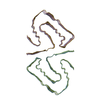
|
|---|---|
| 1 |
|
-要素
| #1: タンパク質 | 分子量: 9649.585 Da / 分子数: 7 / 由来タイプ: 組換発現 / 由来: (組換発現)  |
|---|
-実験情報
-実験
| 実験 | 手法: 電子顕微鏡法 |
|---|---|
| EM実験 | 試料の集合状態: FILAMENT / 3次元再構成法: らせん対称体再構成法 |
- 試料調製
試料調製
| 構成要素 | 名称: Fibril of PI3K-SH3 domains / タイプ: COMPLEX / Entity ID: all / 由来: RECOMBINANT |
|---|---|
| 分子量 | 値: 34.55 kDa/nm / 実験値: NO |
| 由来(天然) | 生物種: |
| 由来(組換発現) | 生物種: |
| 緩衝液 | pH: 2.5 |
| 緩衝液成分 | 濃度: 0.11 mg/mL / 名称: glycin-hydrochloride / 式: C2H6ClNO2 |
| 試料 | 濃度: 1.62 mg/ml / 包埋: NO / シャドウイング: NO / 染色: NO / 凍結: YES |
| 試料支持 | グリッドの材料: COPPER / グリッドのサイズ: 300 divisions/in. / グリッドのタイプ: Quantifoil R1.2/1.3 |
| 急速凍結 | 凍結剤: ETHANE |
- 電子顕微鏡撮影
| 実験機器 |  モデル: Talos Arctica / 画像提供: FEI Company |
|---|---|
| 顕微鏡 | モデル: FEI TECNAI ARCTICA |
| 電子銃 | 電子線源:  FIELD EMISSION GUN / 加速電圧: 200 kV / 照射モード: FLOOD BEAM FIELD EMISSION GUN / 加速電圧: 200 kV / 照射モード: FLOOD BEAM |
| 電子レンズ | モード: BRIGHT FIELD / Cs: 2.7 mm |
| 撮影 | 平均露光時間: 65 sec. / 電子線照射量: 26 e/Å2 / 検出モード: COUNTING フィルム・検出器のモデル: FEI FALCON III (4k x 4k) 実像数: 622 |
| 画像スキャン | 横: 4096 / 縦: 4096 |
- 解析
解析
| EMソフトウェア |
| ||||||||||||||||||||||||||||||||
|---|---|---|---|---|---|---|---|---|---|---|---|---|---|---|---|---|---|---|---|---|---|---|---|---|---|---|---|---|---|---|---|---|---|
| CTF補正 | タイプ: PHASE FLIPPING AND AMPLITUDE CORRECTION | ||||||||||||||||||||||||||||||||
| らせん対称 | 回転角度/サブユニット: 179.436 ° / 軸方向距離/サブユニット: 2.3548 Å / らせん対称軸の対称性: C1 | ||||||||||||||||||||||||||||||||
| 粒子像の選択 | 選択した粒子像数: 103733 / 詳細: Filaments were picked manually in Relion2 | ||||||||||||||||||||||||||||||||
| 3次元再構成 | 解像度: 3.4 Å / 解像度の算出法: FSC 0.143 CUT-OFF / 粒子像の数: 27681 詳細: For the even/odd test, the segment images were split by entire fibrils and refined separately for 25 iterations using the same reference density (low-passed filtered to 20 A). 対称性のタイプ: HELICAL | ||||||||||||||||||||||||||||||||
| 原子モデル構築 | プロトコル: AB INITIO MODEL / 空間: REAL |
