 Movie
Movie Controller

[English] 日本語
 Yorodumi
- PDB-6os9: human Neurotensin Receptor 1 (hNTSR1) - Gi1 Protein Complex in ca... -
+ Open data

- Basic information
| Entry | Database: PDB / ID: 6os9 | |||||||||||||||||||||||||||||||||
|---|---|---|---|---|---|---|---|---|---|---|---|---|---|---|---|---|---|---|---|---|---|---|---|---|---|---|---|---|---|---|---|---|---|---|
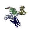
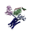
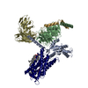
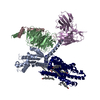
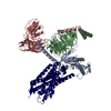
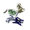
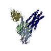
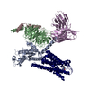
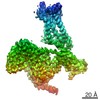
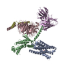
| Title | human Neurotensin Receptor 1 (hNTSR1) - Gi1 Protein Complex in canonical conformation (C state) | |||||||||||||||||||||||||||||||||
 Components Components |
| |||||||||||||||||||||||||||||||||
 Keywords Keywords | SIGNALING PROTEIN / GPCR / G-protein / complex | |||||||||||||||||||||||||||||||||
| Function / homology |  Function and homology information Function and homology informationG protein-coupled neurotensin receptor activity / inositol phosphate catabolic process / symmetric synapse / positive regulation of locomotion / regulation of inositol trisphosphate biosynthetic process / positive regulation of gamma-aminobutyric acid secretion / D-aspartate import across plasma membrane / positive regulation of arachidonate secretion / vocalization behavior / L-glutamate import across plasma membrane ...G protein-coupled neurotensin receptor activity / inositol phosphate catabolic process / symmetric synapse / positive regulation of locomotion / regulation of inositol trisphosphate biosynthetic process / positive regulation of gamma-aminobutyric acid secretion / D-aspartate import across plasma membrane / positive regulation of arachidonate secretion / vocalization behavior / L-glutamate import across plasma membrane / regulation of behavioral fear response / regulation of respiratory gaseous exchange / cAMP biosynthetic process / negative regulation of systemic arterial blood pressure / positive regulation of inhibitory postsynaptic potential / negative regulation of release of sequestered calcium ion into cytosol / response to food / positive regulation of glutamate secretion / regulation of membrane depolarization / response to lipid / temperature homeostasis / positive regulation of inositol phosphate biosynthetic process / response to stress / detection of temperature stimulus involved in sensory perception of pain / conditioned place preference / regulation of eating behavior / adenylate cyclase inhibitor activity / T cell migration / positive regulation of protein localization to cell cortex / positive regulation of relaxation of smooth muscle / Adenylate cyclase inhibitory pathway / D2 dopamine receptor binding / adenylate cyclase-inhibiting serotonin receptor signaling pathway / G protein-coupled serotonin receptor binding / positive regulation of release of sequestered calcium ion into cytosol / cellular response to forskolin / Peptide ligand-binding receptors / regulation of mitotic spindle organization / dendritic shaft / mast cell degranulation / chemokine-mediated signaling pathway / neuropeptide signaling pathway / Regulation of insulin secretion / response to prostaglandin E / positive regulation of cholesterol biosynthetic process / G protein-coupled receptor binding / response to peptide hormone / G protein-coupled receptor activity / cytoplasmic side of plasma membrane / G-protein beta/gamma-subunit complex binding / adenylate cyclase-modulating G protein-coupled receptor signaling pathway / adenylate cyclase-inhibiting G protein-coupled receptor signaling pathway / Olfactory Signaling Pathway / terminal bouton / Activation of the phototransduction cascade / G protein-coupled acetylcholine receptor signaling pathway / G beta:gamma signalling through PLC beta / Presynaptic function of Kainate receptors / Thromboxane signalling through TP receptor / Activation of G protein gated Potassium channels / Inhibition of voltage gated Ca2+ channels via Gbeta/gamma subunits / G-protein activation / Glucagon signaling in metabolic regulation / G beta:gamma signalling through CDC42 / Prostacyclin signalling through prostacyclin receptor / Synthesis, secretion, and inactivation of Glucagon-like Peptide-1 (GLP-1) / GDP binding / G beta:gamma signalling through BTK / photoreceptor disc membrane / ADP signalling through P2Y purinoceptor 12 / Glucagon-type ligand receptors / Sensory perception of sweet, bitter, and umami (glutamate) taste / Adrenaline,noradrenaline inhibits insulin secretion / Vasopressin regulates renal water homeostasis via Aquaporins / Glucagon-like Peptide-1 (GLP1) regulates insulin secretion / G alpha (z) signalling events / cellular response to catecholamine stimulus / ADP signalling through P2Y purinoceptor 1 / G beta:gamma signalling through PI3Kgamma / ADORA2B mediated anti-inflammatory cytokines production / adenylate cyclase-activating dopamine receptor signaling pathway / Cooperation of PDCL (PhLP1) and TRiC/CCT in G-protein beta folding / cellular response to prostaglandin E stimulus / GPER1 signaling / heterotrimeric G-protein complex / Inactivation, recovery and regulation of the phototransduction cascade / G alpha (12/13) signalling events / G-protein beta-subunit binding / extracellular vesicle / Thrombin signalling through proteinase activated receptors (PARs) / signaling receptor complex adaptor activity / adenylate cyclase-activating G protein-coupled receptor signaling pathway / GTPase binding / G protein activity / midbody / chemical synaptic transmission / Ca2+ pathway / cell cortex / High laminar flow shear stress activates signaling by PIEZO1 and PECAM1:CDH5:KDR in endothelial cells / G alpha (i) signalling events Similarity search - Function | |||||||||||||||||||||||||||||||||
| Biological species |  Homo sapiens (human) Homo sapiens (human) synthetic construct (others) | |||||||||||||||||||||||||||||||||
| Method | ELECTRON MICROSCOPY / single particle reconstruction / cryo EM / Resolution: 3 Å | |||||||||||||||||||||||||||||||||
 Authors Authors | Kato, H.E. / Zhang, Y. / Kobilka, B.K. / Skiniotis, G. | |||||||||||||||||||||||||||||||||
| Funding support |  Japan, Japan,  Denmark, Denmark,  United States, 6items United States, 6items
| |||||||||||||||||||||||||||||||||
 Citation Citation | Journal: Nature / Year: 2019 Title: Conformational transitions of a neurotensin receptor 1-G complex. Authors: Hideaki E Kato / Yan Zhang / Hongli Hu / Carl-Mikael Suomivuori / Francois Marie Ngako Kadji / Junken Aoki / Kaavya Krishna Kumar / Rasmus Fonseca / Daniel Hilger / Weijiao Huang / Naomi R ...Authors: Hideaki E Kato / Yan Zhang / Hongli Hu / Carl-Mikael Suomivuori / Francois Marie Ngako Kadji / Junken Aoki / Kaavya Krishna Kumar / Rasmus Fonseca / Daniel Hilger / Weijiao Huang / Naomi R Latorraca / Asuka Inoue / Ron O Dror / Brian K Kobilka / Georgios Skiniotis /  Abstract: Neurotensin receptor 1 (NTSR1) is a G-protein-coupled receptor (GPCR) that engages multiple subtypes of G protein, and is involved in the regulation of blood pressure, body temperature, weight and ...Neurotensin receptor 1 (NTSR1) is a G-protein-coupled receptor (GPCR) that engages multiple subtypes of G protein, and is involved in the regulation of blood pressure, body temperature, weight and the response to pain. Here we present structures of human NTSR1 in complex with the agonist JMV449 and the heterotrimeric G protein, at a resolution of 3 Å. We identify two conformations: a canonical-state complex that is similar to recently reported GPCR-G complexes (in which the nucleotide-binding pocket adopts more flexible conformations that may facilitate nucleotide exchange), and a non-canonical state in which the G protein is rotated by about 45 degrees relative to the receptor and exhibits a more rigid nucleotide-binding pocket. In the non-canonical state, NTSR1 exhibits features of both active and inactive conformations, which suggests that the structure may represent an intermediate form along the activation pathway of G proteins. This structural information, complemented by molecular dynamics simulations and functional studies, provides insights into the complex process of G-protein activation. | |||||||||||||||||||||||||||||||||
| History |
|
- Structure visualization
| Movie |
Movie viewer |
|---|---|
| Structure viewer | Molecule: MolmilJmol/JSmol |
- Downloads & links
-Download
| PDBx/mmCIF format | 6os9.cif.gz | 237.8 KB | Display | PDBx/mmCIF format |
|---|---|---|---|---|
| PDB format | pdb6os9.ent.gz | 179.7 KB | Display | PDB format |
| PDBx/mmJSON format | 6os9.json.gz | Tree view | PDBx/mmJSON format | |
| Others |  Other downloads Other downloads |
-Validation report
| Arichive directory | https://data.pdbj.org/pub/pdb/validation_reports/os/6os9ftp://data.pdbj.org/pub/pdb/validation_reports/os/6os9 | HTTPS FTP |
|---|
-Related structure data
| Related structure data | 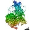 20180MC  6osaC M: map data used to model this data C: citing same article ( |
|---|---|
| Similar structure data |

-Links
 PDBj
PDBj


- Assembly
| Deposited unit | 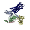
|
|---|---|
| 1 |
|
-Components
-Guanine nucleotide-binding protein ... , 3 types, 3 molecules ABC
| #3: Protein | Mass: 40415.031 Da / Num. of mol.: 1 Source method: isolated from a genetically manipulated source Source: (gene. exp.) Homo sapiens (human) / Gene: GNAI1 / Production host:  Trichoplusia ni (cabbage looper) / References: UniProt: P63096 Trichoplusia ni (cabbage looper) / References: UniProt: P63096 |
|---|---|
| #4: Protein | Mass: 37671.102 Da / Num. of mol.: 1 Source method: isolated from a genetically manipulated source Source: (gene. exp.) Homo sapiens (human) / Gene: GNB1 / Production host: Trichoplusia ni (cabbage looper) / References: UniProt: P62873 |
| #5: Protein | Mass: 7861.143 Da / Num. of mol.: 1 Source method: isolated from a genetically manipulated source Source: (gene. exp.) Homo sapiens (human) / Gene: GNG2 / Production host: Trichoplusia ni (cabbage looper) / References: UniProt: P59768 |
-Protein / Protein/peptide / Antibody , 3 types, 3 molecules RLD
| #1: Protein | Mass: 48396.734 Da / Num. of mol.: 1 / Mutation: A85L Source method: isolated from a genetically manipulated source Source: (gene. exp.) Homo sapiens (human) / Gene: NTSR1, NTRR / Production host:  Spodoptera frugiperda (fall armyworm) / References: UniProt: P30989 Spodoptera frugiperda (fall armyworm) / References: UniProt: P30989 |
|---|---|
| #2: Protein/peptide | Mass: 762.979 Da / Num. of mol.: 1 / Source method: obtained synthetically / Details: JMV449 from Tocris Bioscience. / Source: (synth.) synthetic construct (others) |
| #6: Antibody | Mass: 27784.896 Da / Num. of mol.: 1 Source method: isolated from a genetically manipulated source Source: (gene. exp.) Trichoplusia ni (cabbage looper) |
-Details
| Has protein modification | Y |
|---|
-Experimental details
-Experiment
| Experiment | Method: ELECTRON MICROSCOPY |
|---|---|
| EM experiment | Aggregation state: PARTICLE / 3D reconstruction method: single particle reconstruction |
- Sample preparation
| Component | Name: hNTSR1-Gi1 complex in canonical conformation (C state) Type: COMPLEX / Entity ID: all / Source: RECOMBINANT |
|---|---|
| Molecular weight | Experimental value: NO |
| Source (natural) | Organism: Homo sapiens (human) |
| Source (recombinant) | Organism: Spodoptera frugiperda (fall armyworm) |
| Buffer solution | pH: 7.5 |
| Specimen | Embedding applied: NO / Shadowing applied: NO / Staining applied: NO / Vitrification applied: YES |
| Vitrification | Cryogen name: ETHANE |
- Electron microscopy imaging
| Experimental equipment |  Model: Titan Krios / Image courtesy: FEI Company |
|---|---|
| Microscopy | Model: FEI TITAN KRIOS |
| Electron gun | Electron source:  FIELD EMISSION GUN / Accelerating voltage: 300 kV / Illumination mode: FLOOD BEAM FIELD EMISSION GUN / Accelerating voltage: 300 kV / Illumination mode: FLOOD BEAM |
| Electron lens | Mode: BRIGHT FIELD |
| Image recording | Average exposure time: 10 sec. / Electron dose: 75 e/Å2 / Detector mode: COUNTING / Film or detector model: GATAN K2 SUMMIT (4k x 4k) |
- Processing
| Software | Name: PHENIX / Version: 1.13_2998: / Classification: refinement | ||||||||||||||||||||||||
|---|---|---|---|---|---|---|---|---|---|---|---|---|---|---|---|---|---|---|---|---|---|---|---|---|---|
| EM software | Name: PHENIX / Category: model refinement | ||||||||||||||||||||||||
| CTF correction | Type: PHASE FLIPPING AND AMPLITUDE CORRECTION | ||||||||||||||||||||||||
| 3D reconstruction | Resolution: 3 Å / Resolution method: FSC 0.143 CUT-OFF / Num. of particles: 163333 / Symmetry type: POINT | ||||||||||||||||||||||||
| Refine LS restraints |
|
