 Movie
Movie Controller


+ Open data

- Basic information
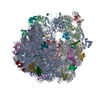
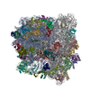
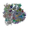
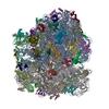
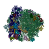
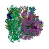
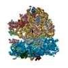
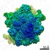
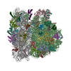
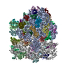
| Entry | Database: PDB / ID: 6o9j | |||||||||||||||
|---|---|---|---|---|---|---|---|---|---|---|---|---|---|---|---|---|
| Title | 70S Elongation Competent Ribosome | |||||||||||||||
 Components Components |
| |||||||||||||||
 Keywords Keywords | RIBOSOME / 70S P/P-tRNA | |||||||||||||||
| Function / homology |  Function and homology information Function and homology informationstringent response / negative regulation of cytoplasmic translational initiation / transcription antitermination factor activity, RNA binding / ornithine decarboxylase inhibitor activity / misfolded RNA binding / Group I intron splicing / RNA folding / translational termination / transcriptional attenuation / endoribonuclease inhibitor activity ...stringent response / negative regulation of cytoplasmic translational initiation / transcription antitermination factor activity, RNA binding / ornithine decarboxylase inhibitor activity / misfolded RNA binding / Group I intron splicing / RNA folding / translational termination / transcriptional attenuation / endoribonuclease inhibitor activity / positive regulation of ribosome biogenesis / RNA-binding transcription regulator activity / four-way junction DNA binding / negative regulation of cytoplasmic translation / regulation of mRNA stability / DnaA-L2 complex / translation repressor activity / negative regulation of translational initiation / negative regulation of DNA-templated DNA replication initiation / mRNA regulatory element binding translation repressor activity / positive regulation of RNA splicing / regulation of DNA-templated transcription elongation / response to reactive oxygen species / transcription elongation factor complex / cytosolic ribosome assembly / ribosome assembly / assembly of large subunit precursor of preribosome / transcription antitermination / DNA endonuclease activity / regulation of cell growth / translational initiation / DNA-templated transcription termination / response to radiation / maintenance of translational fidelity / mRNA 5'-UTR binding / regulation of translation / large ribosomal subunit / transferase activity / ribosomal small subunit assembly / ribosome binding / ribosome biogenesis / ribosomal small subunit biogenesis / 5S rRNA binding / ribosomal large subunit assembly / small ribosomal subunit / small ribosomal subunit rRNA binding / cytosolic small ribosomal subunit / large ribosomal subunit rRNA binding / cytosolic large ribosomal subunit / cytoplasmic translation / tRNA binding / negative regulation of translation / rRNA binding / structural constituent of ribosome / ribosome / translation / ribonucleoprotein complex / response to antibiotic / negative regulation of DNA-templated transcription / hydrolase activity / mRNA binding / DNA binding / RNA binding / zinc ion binding / membrane / metal ion binding / cytosol / cytoplasm Similarity search - Function | |||||||||||||||
| Biological species |  | |||||||||||||||
| Method | ELECTRON MICROSCOPY / single particle reconstruction / cryo EM / Resolution: 3.9 Å | |||||||||||||||
 Authors Authors | Frank, J. / Gonzalez Jr., R.L. / Kaledhonkar, S. / Fu, Z. / Caban, K. / Li, W. / Chen, B. / Sun, M. | |||||||||||||||
| Funding support |  United States, 4items United States, 4items
| |||||||||||||||
 Citation Citation | Journal: Nature / Year: 2019 Title: Late steps in bacterial translation initiation visualized using time-resolved cryo-EM. Authors: Sandip Kaledhonkar / Ziao Fu / Kelvin Caban / Wen Li / Bo Chen / Ming Sun / Ruben L Gonzalez / Joachim Frank / Abstract: The initiation of bacterial translation involves the tightly regulated joining of the 50S ribosomal subunit to an initiator transfer RNA (fMet-tRNA)-containing 30S ribosomal initiation complex to ...The initiation of bacterial translation involves the tightly regulated joining of the 50S ribosomal subunit to an initiator transfer RNA (fMet-tRNA)-containing 30S ribosomal initiation complex to form a 70S initiation complex, which subsequently matures into a 70S elongation-competent complex. Rapid and accurate formation of the 70S initiation complex is promoted by initiation factors, which must dissociate from the 30S initiation complex before the resulting 70S elongation-competent complex can begin the elongation of translation. Although comparisons of the structures of the 30S and 70S initiation complexes have revealed that the ribosome, initiation factors and fMet-tRNA can acquire different conformations in these complexes, the timing of conformational changes during formation of the 70S initiation complex, the structures of any intermediates formed during these rearrangements, and the contributions that these dynamics might make to the mechanism and regulation of initiation remain unknown. Moreover, the absence of a structure of the 70S elongation-competent complex formed via an initiation-factor-catalysed reaction has precluded an understanding of the rearrangements to the ribosome, initiation factors and fMet-tRNA that occur during maturation of a 70S initiation complex into a 70S elongation-competent complex. Here, using time-resolved cryogenic electron microscopy, we report the near-atomic-resolution view of how a time-ordered series of conformational changes drive and regulate subunit joining, initiation factor dissociation and fMet-tRNA positioning during formation of the 70S elongation-competent complex. Our results demonstrate the power of time-resolved cryogenic electron microscopy to determine how a time-ordered series of conformational changes contribute to the mechanism and regulation of one of the most fundamental processes in biology. | |||||||||||||||
| History |
|
- Structure visualization
| Movie |
Movie viewer |
|---|---|
| Structure viewer | Molecule: MolmilJmol/JSmol |
- Downloads & links
-Download
| PDBx/mmCIF format | 6o9j.cif.gz | 3.2 MB | Display | PDBx/mmCIF format |
|---|---|---|---|---|
| PDB format | pdb6o9j.ent.gz | Display | PDB format | |
| PDBx/mmJSON format | 6o9j.json.gz | Tree view | PDBx/mmJSON format | |
| Others |  Other downloads Other downloads |
-Validation report
| Arichive directory | https://data.pdbj.org/pub/pdb/validation_reports/o9/6o9jftp://data.pdbj.org/pub/pdb/validation_reports/o9/6o9j | HTTPS FTP |
|---|
-Related structure data
| Related structure data | 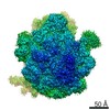 0661MC  0643C 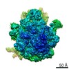 0662C  6o7kC  6o9kC M: map data used to model this data C: citing same article ( |
|---|---|
| Similar structure data |
-Links
 PDBj
PDBj


- Assembly
| Deposited unit | 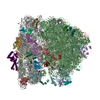
|
|---|---|
| 1 |
|
-Components
-RNA chain , 4 types, 4 molecules ABav
| #1: RNA chain | Mass: 37848.555 Da / Num. of mol.: 1 / Source method: isolated from a natural source / Source: (natural) |
|---|---|
| #2: RNA chain | Mass: 921273.375 Da / Num. of mol.: 1 / Source method: isolated from a natural source / Source: (natural) |
| #32: RNA chain | Mass: 495927.719 Da / Num. of mol.: 1 / Source method: isolated from a natural source / Source: (natural) |
| #53: RNA chain | Mass: 24485.539 Da / Num. of mol.: 1 / Source method: isolated from a natural source / Source: (natural) |
+50S ribosomal protein ... , 29 types, 29 molecules VCDEFGHJKLMNOPQRSTUWXYZ123456
-30S ribosomal protein ... , 20 types, 20 molecules cdefghijklmnopqrst7u
| #33: Protein | Mass: 23078.785 Da / Num. of mol.: 1 / Source method: isolated from a natural source / Source: (natural) |
|---|---|
| #34: Protein | Mass: 23383.002 Da / Num. of mol.: 1 / Source method: isolated from a natural source / Source: (natural) |
| #35: Protein | Mass: 15804.282 Da / Num. of mol.: 1 / Source method: isolated from a natural source / Source: (natural) |
| #36: Protein | Mass: 11669.371 Da / Num. of mol.: 1 / Source method: isolated from a natural source / Source: (natural) |
| #37: Protein | Mass: 16764.406 Da / Num. of mol.: 1 / Source method: isolated from a natural source / Source: (natural) |
| #38: Protein | Mass: 14015.361 Da / Num. of mol.: 1 / Source method: isolated from a natural source / Source: (natural) |
| #39: Protein | Mass: 14554.882 Da / Num. of mol.: 1 / Source method: isolated from a natural source / Source: (natural) |
| #40: Protein | Mass: 11196.988 Da / Num. of mol.: 1 / Source method: isolated from a natural source / Source: (natural) |
| #41: Protein | Mass: 12487.200 Da / Num. of mol.: 1 / Source method: isolated from a natural source / Source: (natural) |
| #42: Protein | Mass: 13636.961 Da / Num. of mol.: 1 / Source method: isolated from a natural source / Source: (natural) |
| #43: Protein | Mass: 12625.753 Da / Num. of mol.: 1 / Source method: isolated from a natural source / Source: (natural) |
| #44: Protein | Mass: 11489.390 Da / Num. of mol.: 1 / Source method: isolated from a natural source / Source: (natural) |
| #45: Protein | Mass: 10188.687 Da / Num. of mol.: 1 / Source method: isolated from a natural source / Source: (natural) |
| #46: Protein | Mass: 9207.572 Da / Num. of mol.: 1 / Source method: isolated from a natural source / Source: (natural) |
| #47: Protein | Mass: 9263.946 Da / Num. of mol.: 1 / Source method: isolated from a natural source / Source: (natural) |
| #48: Protein | Mass: 6466.477 Da / Num. of mol.: 1 / Source method: isolated from a natural source / Source: (natural) |
| #49: Protein | Mass: 9057.626 Da / Num. of mol.: 1 / Source method: isolated from a natural source / Source: (natural) |
| #50: Protein | Mass: 9506.190 Da / Num. of mol.: 1 / Source method: isolated from a natural source / Source: (natural) |
| #51: Protein | Mass: 24253.943 Da / Num. of mol.: 1 / Source method: isolated from a natural source / Source: (natural) |
| #52: Protein | Mass: 6067.081 Da / Num. of mol.: 1 / Source method: isolated from a natural source / Source: (natural) |
-Experimental details
-Experiment
| Experiment | Method: ELECTRON MICROSCOPY |
|---|---|
| EM experiment | Aggregation state: PARTICLE / 3D reconstruction method: single particle reconstruction |
- Sample preparation
| Component | Name: 70S elongation competent ribosome / Type: RIBOSOME / Entity ID: all / Source: NATURAL |
|---|---|
| Source (natural) | Organism: |
| Buffer solution | pH: 7.2 |
| Specimen | Embedding applied: NO / Shadowing applied: NO / Staining applied: NO / Vitrification applied: YES |
| Specimen support | Details: unspecified |
| Vitrification | Cryogen name: ETHANE |
- Electron microscopy imaging
| Experimental equipment |  Model: Tecnai F30 / Image courtesy: FEI Company |
|---|---|
| Microscopy | Model: FEI TECNAI F30 |
| Electron gun | Electron source:  FIELD EMISSION GUN / Accelerating voltage: 300 kV / Illumination mode: OTHER FIELD EMISSION GUN / Accelerating voltage: 300 kV / Illumination mode: OTHER |
| Electron lens | Mode: DARK FIELD |
| Image recording | Electron dose: 8 e/Å2 / Film or detector model: GATAN K2 SUMMIT (4k x 4k) |
- Processing
| CTF correction | Type: PHASE FLIPPING AND AMPLITUDE CORRECTION |
|---|---|
| 3D reconstruction | Resolution: 3.9 Å / Resolution method: FSC 0.143 CUT-OFF / Num. of particles: 46042 / Symmetry type: POINT |
