 Movie
Movie Controller

[English] 日本語
 Yorodumi
- PDB-6nsj: CryoEM structure of Helicobacter pylori urea channel in closed state -
+ Open data

- Basic information
| Entry | Database: PDB / ID: 6nsj | |||||||||
|---|---|---|---|---|---|---|---|---|---|---|
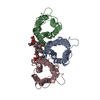
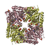
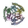
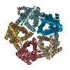
| Title | CryoEM structure of Helicobacter pylori urea channel in closed state | |||||||||
 Components Components | Acid-activated urea channel | |||||||||
 Keywords Keywords | TRANSPORT PROTEIN / Helicobacter pylori / urea channel / closed state | |||||||||
| Function / homology |  Function and homology information Function and homology information | |||||||||
| Biological species |   Helicobacter pylori (bacteria) Helicobacter pylori (bacteria) | |||||||||
| Method | ELECTRON MICROSCOPY / single particle reconstruction / cryo EM / Resolution: 2.7 Å | |||||||||
 Authors Authors | Cui, Y.X. / Zhou, K. / Strugatsky, D. / Wen, Y. / Sachs, G. / Munson, K. / Zhou, Z.H. | |||||||||
| Funding support |  United States, 2items United States, 2items
| |||||||||
 Citation Citation | Journal: Sci Adv / Year: 2019 Title: pH-dependent gating mechanism of the urea channel revealed by cryo-EM. Authors: Yanxiang Cui / Kang Zhou / David Strugatsky / Yi Wen / George Sachs / Z Hong Zhou / Keith Munson / Abstract: The urea channel of (UreI) is an ideal drug target for preventing gastric cancer but incomplete understanding of its gating mechanism has hampered development of inhibitors for the eradication of . ...The urea channel of (UreI) is an ideal drug target for preventing gastric cancer but incomplete understanding of its gating mechanism has hampered development of inhibitors for the eradication of . Here, we present the cryo-EM structures of UreI in closed and open conformations, both at a resolution of 2.7 Å. Our hexameric structures of this small membrane protein (~21 kDa/protomer) resolve its periplasmic loops and carboxyl terminus that close and open the channel, and define a gating mechanism that is pH dependent and requires cooperativity between protomers in the hexamer. Gating is further associated with well-resolved changes in the channel-lining residues that modify the shape and length of the urea pore. Site-specific mutations in the periplasmic domain and urea pore identified key residues important for channel function. Drugs blocking the urea pore based on our structures should lead to a new strategy for eradication. | |||||||||
| History |
|
- Structure visualization
| Movie |
Movie viewer |
|---|---|
| Structure viewer | Molecule: MolmilJmol/JSmol |
- Downloads & links
-Download
| PDBx/mmCIF format | 6nsj.cif.gz | 205.6 KB | Display | PDBx/mmCIF format |
|---|---|---|---|---|
| PDB format | pdb6nsj.ent.gz | 165.7 KB | Display | PDB format |
| PDBx/mmJSON format | 6nsj.json.gz | Tree view | PDBx/mmJSON format | |
| Others |  Other downloads Other downloads |
-Validation report
| Arichive directory | https://data.pdbj.org/pub/pdb/validation_reports/ns/6nsjftp://data.pdbj.org/pub/pdb/validation_reports/ns/6nsj | HTTPS FTP |
|---|
-Related structure data
| Related structure data |  0498MC 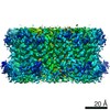 0499C 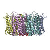 6nskC M: map data used to model this data C: citing same article ( |
|---|---|
| Similar structure data |


-Links
 PDBj
PDBj
- Assembly
| Deposited unit | 
|
|---|---|
| 1 |
|
-Components
| #1: Protein | Mass: 22533.260 Da / Num. of mol.: 6 Source method: isolated from a genetically manipulated source Source: (gene. exp.) Helicobacter pylori (strain J99 / ATCC 700824) (bacteria)Strain: J99 / ATCC 700824 / Gene: ureI, jhp_0066 / Production host: #2: Chemical | ChemComp-XP4 /   Mass: 591.777 Da / Num. of mol.: 18 / Source method: obtained synthetically / Formula: C31H60O8P / Comment: DMPA, phospholipid*YM Mass: 591.777 Da / Num. of mol.: 18 / Source method: obtained synthetically / Formula: C31H60O8P / Comment: DMPA, phospholipid*YM |
|---|
-Experimental details
-Experiment
| Experiment | Method: ELECTRON MICROSCOPY |
|---|---|
| EM experiment | Aggregation state: 2D ARRAY / 3D reconstruction method: single particle reconstruction |
- Sample preparation
| Component | Name: urea channel of Helicobacter pylori / Type: ORGANELLE OR CELLULAR COMPONENT / Entity ID: #1 / Source: RECOMBINANT | ||||||||||||
|---|---|---|---|---|---|---|---|---|---|---|---|---|---|
| Molecular weight | Value: 0.02 MDa / Experimental value: YES | ||||||||||||
| Source (natural) | Organism: Helicobacter pylori (bacteria) / Strain: J99 | ||||||||||||
| Source (recombinant) | Organism: | ||||||||||||
| Buffer solution | pH: 7 | ||||||||||||
| Buffer component |
| ||||||||||||
| Specimen | Conc.: 2 mg/ml / Embedding applied: NO / Shadowing applied: NO / Staining applied: NO / Vitrification applied: YES | ||||||||||||
| Specimen support | Grid material: COPPER / Grid mesh size: 300 divisions/in. / Grid type: Quantifoil R2/1 | ||||||||||||
| Vitrification | Instrument: FEI VITROBOT MARK IV / Cryogen name: ETHANE / Humidity: 100 % / Chamber temperature: 277.15 K |
- Electron microscopy imaging
| Experimental equipment |  Model: Titan Krios / Image courtesy: FEI Company |
|---|---|
| Microscopy | Model: FEI TITAN KRIOS |
| Electron gun | Electron source:  FIELD EMISSION GUN / Accelerating voltage: 300 kV / Illumination mode: FLOOD BEAM FIELD EMISSION GUN / Accelerating voltage: 300 kV / Illumination mode: FLOOD BEAM |
| Electron lens | Mode: BRIGHT FIELD / Nominal magnification: 130000 X / Calibrated magnification: 46730 X / Nominal defocus max: 2400 nm / Nominal defocus min: 1400 nm / Calibrated defocus min: 1200 nm / Calibrated defocus max: 2600 nm / Cs: 2.7 mm / C2 aperture diameter: 70 µm / Alignment procedure: COMA FREE |
| Specimen holder | Cryogen: NITROGEN / Specimen holder model: FEI TITAN KRIOS AUTOGRID HOLDER / Temperature (max): 100 K / Temperature (min): 98 K |
| Image recording | Average exposure time: 15 sec. / Electron dose: 72 e/Å2 / Detector mode: SUPER-RESOLUTION / Film or detector model: GATAN K2 SUMMIT (4k x 4k) / Num. of grids imaged: 1 / Num. of real images: 4026 |
| EM imaging optics | Energyfilter name: GIF Quantum LS / Energyfilter slit width: 20 eV |
| Image scans | Sampling size: 5 µm / Width: 3710 / Height: 3838 / Movie frames/image: 100 / Used frames/image: 2-99 |
- Processing
| EM software |
| ||||||||||||||||||||||||||||||||||||||||
|---|---|---|---|---|---|---|---|---|---|---|---|---|---|---|---|---|---|---|---|---|---|---|---|---|---|---|---|---|---|---|---|---|---|---|---|---|---|---|---|---|---|
| CTF correction | Type: NONE | ||||||||||||||||||||||||||||||||||||||||
| Particle selection | Num. of particles selected: 1221897 | ||||||||||||||||||||||||||||||||||||||||
| Symmetry | Point symmetry: C6 (6 fold cyclic) | ||||||||||||||||||||||||||||||||||||||||
| 3D reconstruction | Resolution: 2.7 Å / Resolution method: FSC 0.143 CUT-OFF / Num. of particles: 393930 / Algorithm: FOURIER SPACE / Num. of class averages: 3 / Symmetry type: POINT | ||||||||||||||||||||||||||||||||||||||||
| Atomic model building | B value: 71.9 / Protocol: RIGID BODY FIT / Space: REAL | ||||||||||||||||||||||||||||||||||||||||
| Atomic model building | PDB-ID: 3UX4 Accession code: 3UX4 / Source name: PDB / Type: experimental model |

